El-Shenawee Magda, Vohra Nagma, Bowman Tyler, Bailey Keith
Department of Electrical Engineering, University of Arkansas, Fayetteville, USA.
Oklahoma Animal Disease Diagnostic Laboratory, Oklahoma State University, Stillwater, Oklahoma, USA.
Biomed Spectrosc Imaging. 2019;8(1-2):1-9. doi: 10.3233/bsi-190187. Epub 2019 Jul 9.
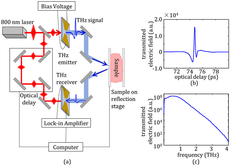
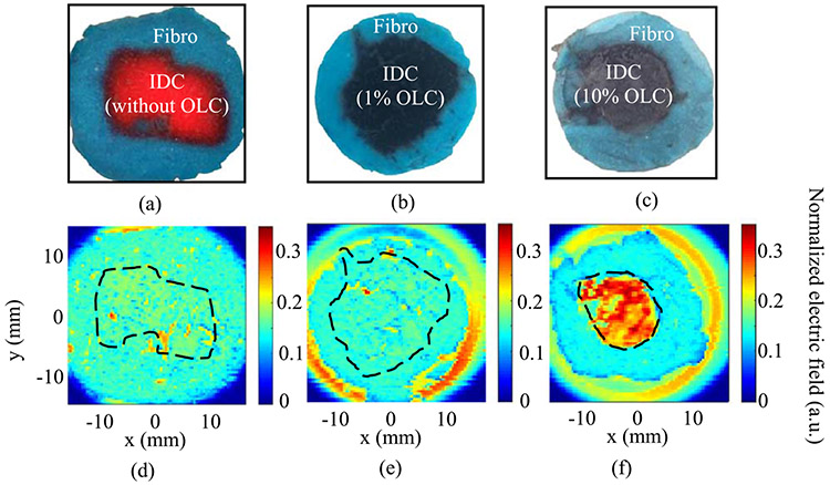
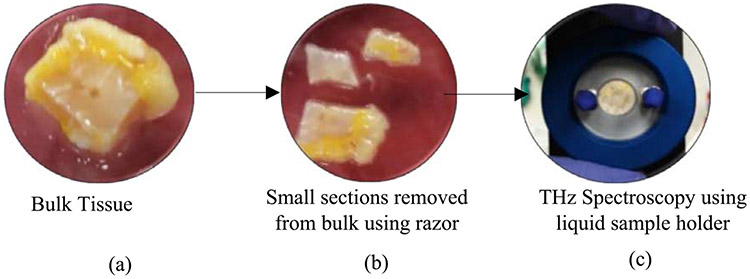
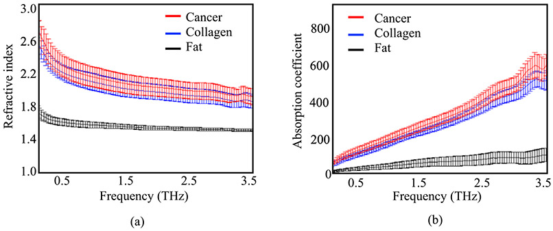
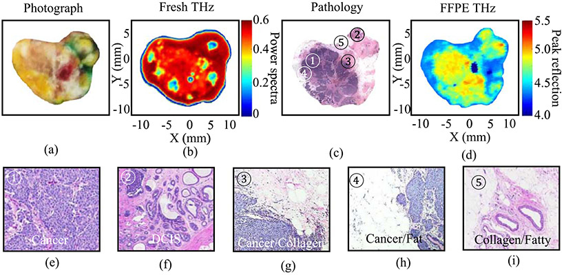
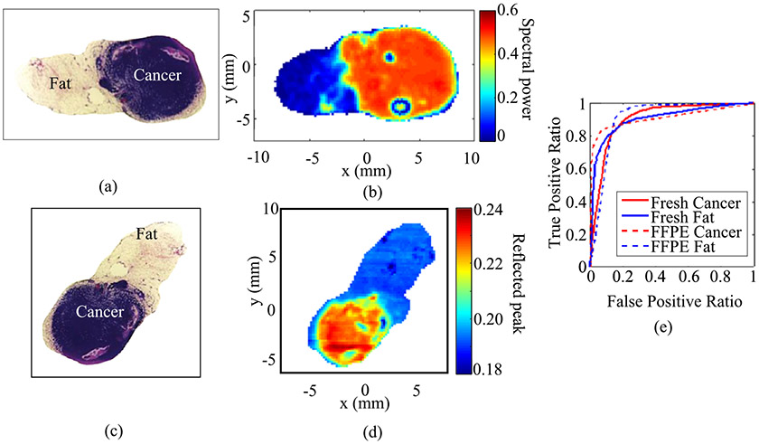
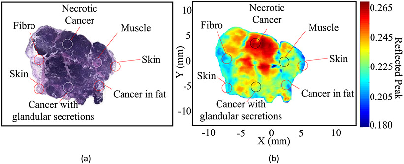
Terahertz imaging and spectroscopy has demonstrated a potential for differentiating tissue types of excised breast cancer tumors. Pulsed terahertz technology provides a broadband frequency range from 0.1 THz to 4 THz for detecting cancerous tissue. Tumor tissue types of interest include cancer typically manifested as infiltrating ductal or lobular carcinomas, fibro-glandular (healthy connective tissues) and fat. In this work, images of breast tumors excised from human and animal models are reviewed. In addition to alternate fresh tissues, breast cancer tissue phantoms are developed to further evaluate terahertz imaging and the potential use of contrast agents. Terahertz results are successfully validated with pathology images, showing strong differentiation between cancerous and healthy tissues for all freshly excised tissues and types. The advantages, challenges and limitations of THz imaging of breast cancer are discussed.
太赫兹成像与光谱技术已展现出区分切除的乳腺癌肿瘤组织类型的潜力。脉冲太赫兹技术提供了从0.1太赫兹到4太赫兹的宽带频率范围,用于检测癌组织。感兴趣的肿瘤组织类型包括通常表现为浸润性导管癌或小叶癌的癌症、纤维腺组织(健康结缔组织)和脂肪。在这项工作中,对从人类和动物模型切除的乳腺肿瘤图像进行了回顾。除了新鲜组织外,还制作了乳腺癌组织模型,以进一步评估太赫兹成像以及造影剂的潜在用途。太赫兹成像结果已通过病理图像成功验证,显示所有新鲜切除的组织及其类型的癌组织与健康组织之间有明显区分。讨论了乳腺癌太赫兹成像的优势、挑战和局限性。