.
,.
Invest Ophthalmol Vis Sci. 2020 Jun 3;61(6):49. doi: 10.1167/iovs.61.6.49.
We hypothesize that patients with type 1 diabetes (T1D) may have abnormal retinal vascular responses before diabetic retinopathy (DR) is clinically evident. Optical coherence tomography angiography (OCTA) was used to dynamically assess the retinal microvasculature of diabetic patients with no clinically visible retinopathy.
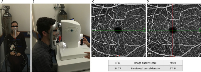
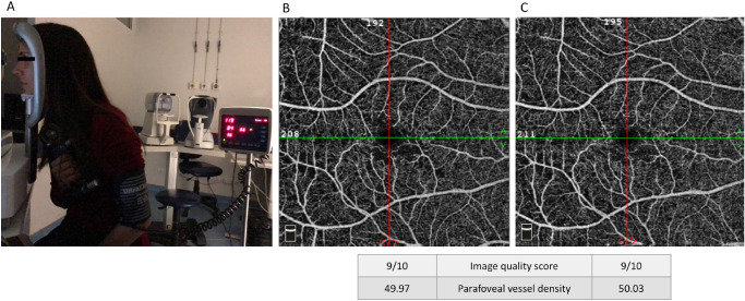
Controlled nonrandomized interventional study. The studied population included 48 eyes of 24 T1D patients and 24 demographically similar healthy volunteers. A commercial OCTA device (AngioVue) was used, and two tests were applied: (1) the hypoxia challenge test (HCT) and (2) the handgrip test to induce a vasodilatory or vasoconstrictive response, respectively. The HCT is a standardized test that creates a mild hypoxic environment equivalent to a flight cabin. The handgrip test (i.e., isometric exercise) induces a sympathetic autonomic response. Changes in the parafoveal superficial and deep capillary plexuses in both tests were compared in each group. Systemic cardiovascular responses were also comparatively evaluated.
In the control cohort, the vessel density of the median parafoveal superficial and deep plexuses increased during hypoxia (F1,23 = 15.69, P < 0.001 and F1,23 = 16.26, P < 0.001, respectively). In the T1D group, this physiological response was not observed in either the superficial or the deep retinal plexuses. Isometric exercise elicited a significant decrease in vessel density in both superficial and deep plexuses in the control group (F1,23 = 27.37, P < 0.0001 and F1,23 = 27.90, P < 0.0001, respectively). In the T1D group, this response was noted only in the deep plexus (F1,23 = 11.04, P < 0.01).
Our work suggests there is an early impairment of the physiological retinal vascular response in patients with T1D without clinical diabetic retinopathy.
我们假设在糖尿病视网膜病变(DR)临床明显之前,1 型糖尿病(T1D)患者可能存在视网膜血管反应异常。本研究采用光学相干断层扫描血管造影(OCTA)对无临床可见视网膜病变的糖尿病患者的视网膜微血管进行动态评估。
对照非随机干预研究。研究人群包括 24 例 T1D 患者的 48 只眼和 24 名年龄匹配的健康志愿者。使用商用 OCTA 设备(AngioVue),并进行两项测试:(1)缺氧挑战测试(HCT)和(2)握力测试,分别诱导血管舒张或收缩反应。HCT 是一种标准化测试,可营造相当于飞行客舱的轻度低氧环境。握力测试(即等长运动)可引起交感自主神经反应。在两组中比较了两种测试中黄斑旁浅层和深层毛细血管丛的变化。还比较了系统心血管反应。
在对照组中,缺氧时中央黄斑旁浅层和深层毛细血管丛的血管密度增加(F1,23 = 15.69,P <0.001 和 F1,23 = 16.26,P <0.001)。在 T1D 组中,浅层和深层视网膜丛均未观察到这种生理反应。等长运动引起对照组浅层和深层毛细血管丛的血管密度显著降低(F1,23 = 27.37,P <0.0001 和 F1,23 = 27.90,P <0.0001)。在 T1D 组中,仅在深层丛中观察到这种反应(F1,23 = 11.04,P <0.01)。
我们的研究结果表明,在无临床糖尿病视网膜病变的 T1D 患者中,视网膜血管的生理反应存在早期损害。