Department of Anatomy and Histology, Faculty of Vet. Medicine, Assiut University, Assiut, Egypt.
Sci Rep. 2020 Jun 23;10(1):10154. doi: 10.1038/s41598-020-67103-5.
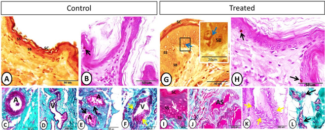
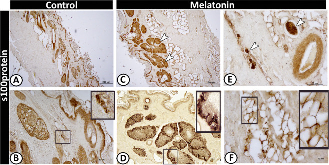
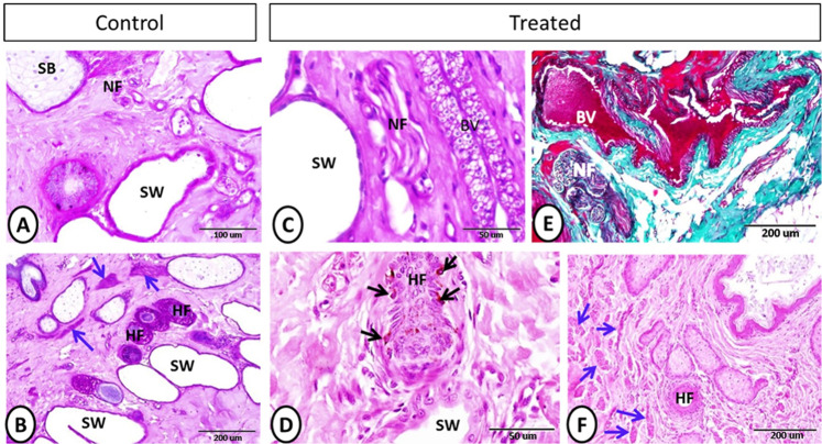
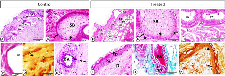
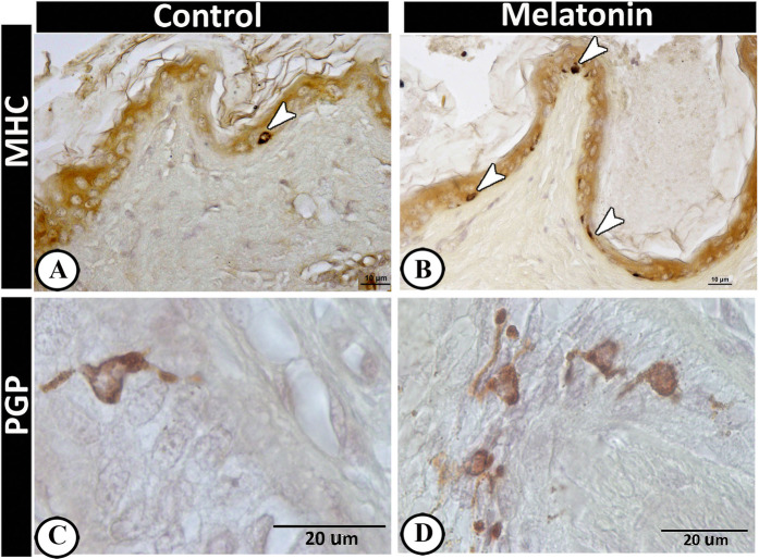
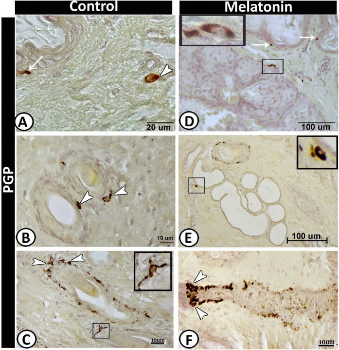
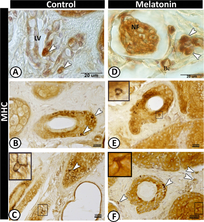
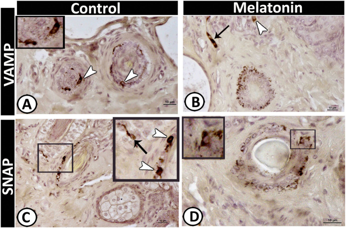
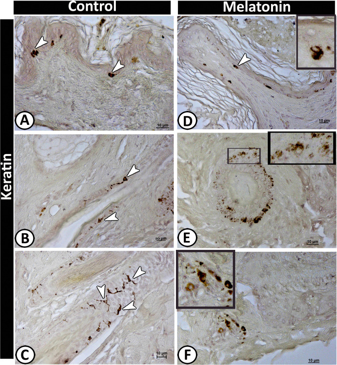
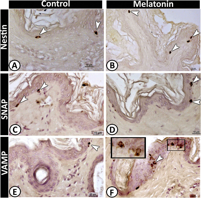
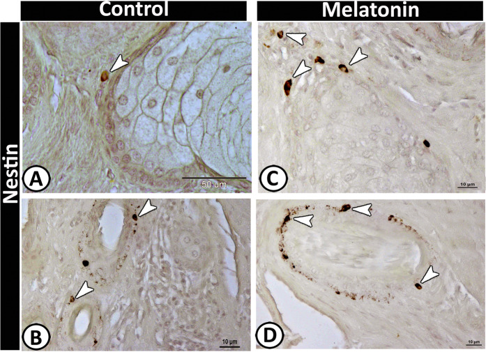
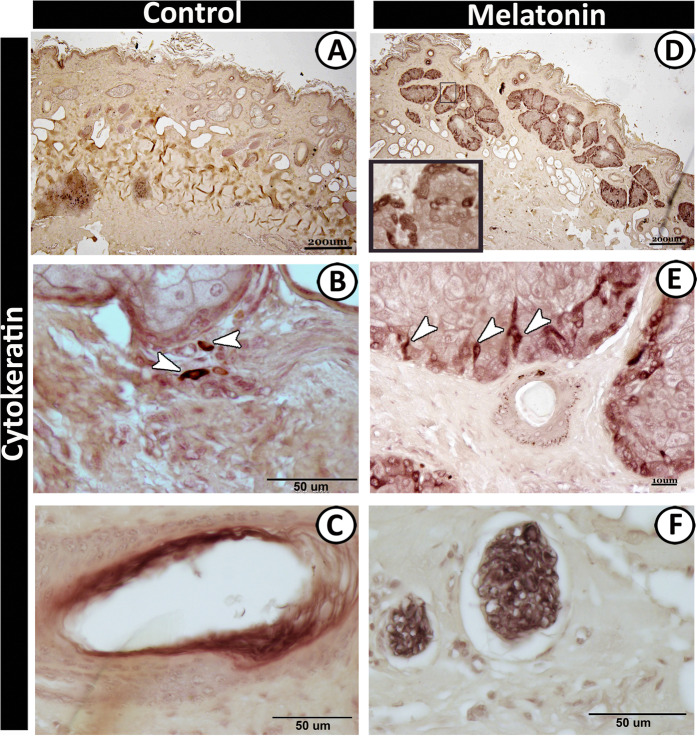
Fifteen adult Soay rams were employed in this study to investigate the effect of melatonin on the scrotal skin using histological, histochemical, and morphometrical analysis. The results revealed that the melatonin treated group showed a significant increase in the thickness of the epidermis, the cross-sectional area of blood capillaries and nerve fibers compared with the control one. In addition, obvious hypertrophy and hyperplasia were detected in the sebaceous glands in association with a significant increase in the number and diameter of apocrine sweat glands with well-developed secretory activity. S100 protein and cytokeratin-19 strongly stained the basal cells of sebaceous glands in the melatonin treated group incomparable to the control group. Moreover, the nerve fibers were intensively immunoreacted for S100 and cytokeratin proteins in the melatonin treated group in contrast to the control one. A high number of telocytes (TCs) could be identified in the treated group around the nerve fibers and blood vessels in the dermis. The number of Langerhans cells showed a significant increase in the melatonin groups that were identified by MHC II and PGP 9.5 within the epidermal layer. Furthermore, a significant increase in the number of dendritic cells was identified in the melatonin group, which were distributed within the dermis, around hair follicles, sebaceous glands, and sweat glands and were strongly expressed PGP-9.5, MHC-II, VAMP, SNAP, keratin-5, and cytokeratin-19 immunoreactivity. Notably, Merkel cells showed a significant increase in the number in the melatonin group that could be stained against nestin, SNAP, and VAMP. On the other hand, the secretory granules in sweat glands were exhibited a strong positive reactivity for synaptophysin in melatonin group. The current study showed that the administration of melatonin induced a stimulatory effect on keratinocytes, non-keratinocytes, sebaceous and sweat glands, hair follicles, as well as the vascular, neuronal, and cellular constituents of the dermis.
本研究采用 15 只成年斯澳绵羊,通过组织学、组织化学和形态计量学分析,探讨褪黑素对阴囊皮肤的影响。结果显示,与对照组相比,褪黑素处理组的表皮厚度、毛细血管和神经纤维的横截面积显著增加。此外,还发现皮脂腺明显肥大和增生,与顶泌汗腺数量和直径的显著增加以及发达的分泌活性有关。S100 蛋白和细胞角蛋白-19 强烈染色褪黑素处理组的皮脂腺基底细胞,而对照组则没有。此外,与对照组相比,褪黑素处理组的神经纤维对 S100 和细胞角蛋白蛋白的免疫反应更为强烈。真皮中神经纤维周围和血管周围可以识别到大量的间质细胞(TCs)。褪黑素组的郎格汉斯细胞数量显著增加,通过 MHC II 和 PGP 9.5 在表皮层中鉴定。此外,褪黑素组中树突状细胞的数量也显著增加,这些细胞分布在真皮中,围绕毛囊、皮脂腺和汗腺,并强烈表达 PGP-9.5、MHC-II、VAMP、SNAP、角蛋白-5 和细胞角蛋白-19 免疫反应性。值得注意的是,Merkel 细胞在褪黑素组中的数量显著增加,可以对巢蛋白、SNAP 和 VAMP 进行染色。另一方面,在褪黑素组中,汗腺的分泌颗粒对突触素呈强阳性反应。本研究表明,褪黑素的给药对角质形成细胞、非角质形成细胞、皮脂腺和汗腺、毛囊以及真皮的血管、神经元和细胞成分具有刺激作用。