• 文献检索
  • 文档翻译
  • 深度研究
  • 学术资讯
  • Suppr Zotero 插件Zotero 插件
  • 邀请有礼
  • 套餐&价格
  • 历史记录
应用&插件
Suppr Zotero 插件Zotero 插件浏览器插件Mac 客户端Windows 客户端微信小程序
定价
高级版会员购买积分包购买API积分包
服务
文献检索文档翻译深度研究API 文档MCP 服务
关于我们
关于 Suppr公司介绍联系我们用户协议隐私条款
关注我们

Suppr 超能文献

核心技术专利:CN118964589B侵权必究
粤ICP备2023148730 号-1Suppr @ 2026

文献检索

告别复杂PubMed语法,用中文像聊天一样搜索,搜遍4000万医学文献。AI智能推荐,让科研检索更轻松。

立即免费搜索

文件翻译

保留排版,准确专业,支持PDF/Word/PPT等文件格式,支持 12+语言互译。

免费翻译文档

深度研究

AI帮你快速写综述,25分钟生成高质量综述,智能提取关键信息,辅助科研写作。

立即免费体验

蛋白质组学分析鉴定硫化镉量子点(CdS QDs)暴露的标志物。

Proteomic Analysis Identifies Markers of Exposure to Cadmium Sulphide Quantum Dots (CdS QDs).

作者信息

Gallo Valentina, Srivastava Vaibhav, Bulone Vincent, Zappettini Andrea, Villani Marco, Marmiroli Nelson, Marmiroli Marta

机构信息

Department of Chemistry, Life Sciences and Environmental Sustainability, University of Parma, 43123 Parma, Italy.

Royal Institute of Technology (KTH), Department of Chemistry, Division of Glycoscience, School of Engineering Sciences in Chemistry, Biotechnology and Health, AlbaNova University Center, SE-106 91 Stockholm, Sweden.

出版信息

Nanomaterials (Basel). 2020 Jun 22;10(6):1214. doi: 10.3390/nano10061214.

DOI:10.3390/nano10061214
PMID:32580447
原文链接:https://pmc.ncbi.nlm.nih.gov/articles/PMC7353101/
Abstract

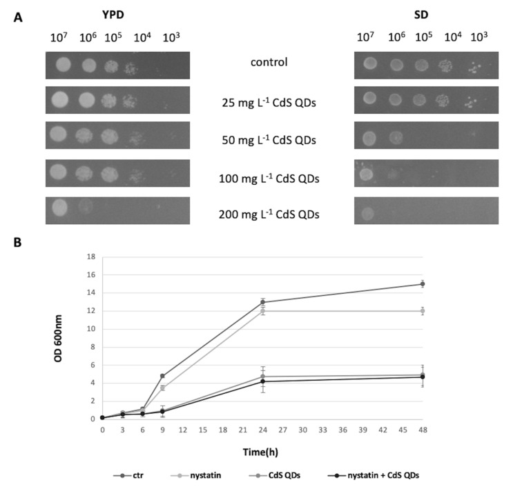
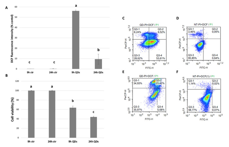
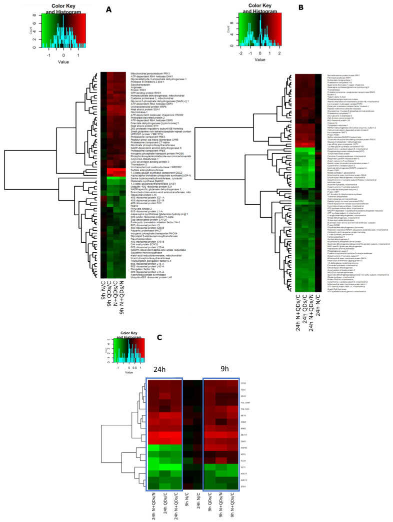
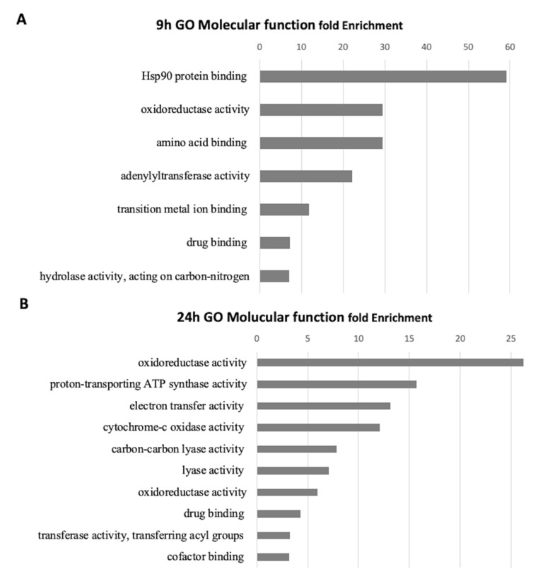
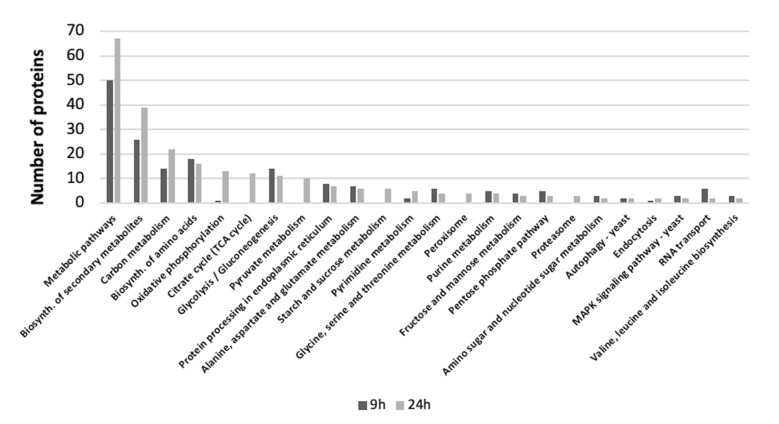
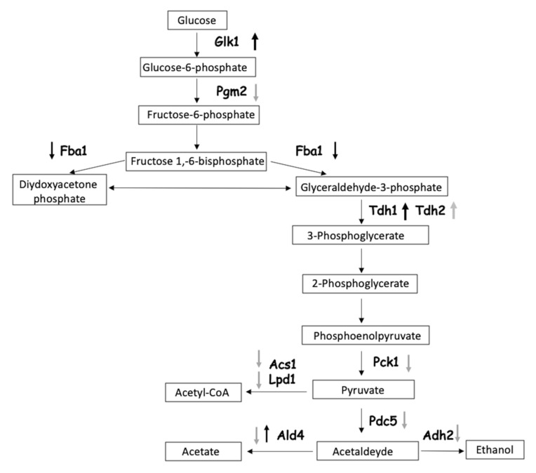
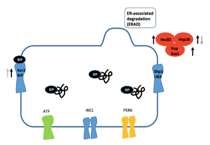
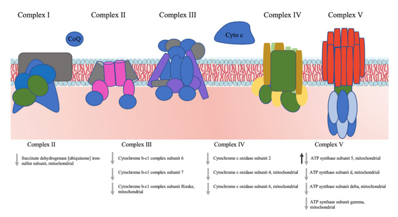
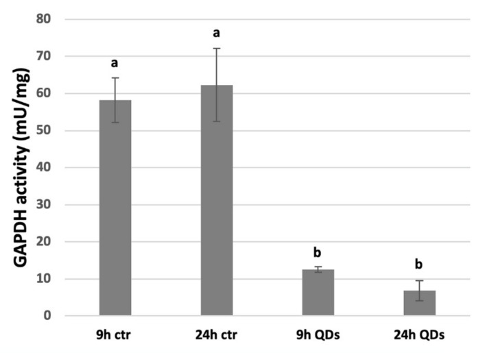
The use of cadmium sulphide quantum dot (CdS QD)-enabled products has become increasingly widespread. The prospect of their release in the environment is raising concerns. Here we have used the yeast model to determine the potential impact of CdS QD nanoparticles on living organisms. Proteomic analyses and cell viability assays performed after 9 h exposure revealed expression of proteins involved in oxidative stress and reduced lethality, respectively, whereas oxidative stress declined, and lethality increased after 24 h incubation in the presence of CdS QDs. Quantitative proteomics using the iTRAQ approach (isobaric tags for relative and absolute quantitation) revealed that key proteins involved in essential biological pathways were differentially regulated over the time course of the experiment. At 9 h, most of the glycolytic functions increased, and the abundance of the number of heat shock proteins increased. This contrasts with the situation at 24 h where glycolytic functions, some heat shock proteins as well as oxidative phosphorylation and ATP synthesis were down-regulated. It can be concluded from our data that cell exposure to CdS QDs provokes a metabolic shift from respiration to fermentation, comparable to the situation reported in some cancer cell lines.

摘要

硫化镉量子点(CdS QD)相关产品的使用日益广泛。其在环境中释放的可能性引发了人们的担忧。在此,我们使用酵母模型来确定CdS QD纳米颗粒对生物体的潜在影响。暴露9小时后进行的蛋白质组学分析和细胞活力测定分别显示了参与氧化应激的蛋白质表达以及致死率降低,而在CdS QDs存在下孵育24小时后,氧化应激下降,致死率增加。使用iTRAQ方法(相对和绝对定量的等压标签)进行的定量蛋白质组学显示,在实验过程中,参与基本生物学途径的关键蛋白质受到不同程度的调控。在9小时时,大多数糖酵解功能增强,热休克蛋白数量的丰度增加。这与24小时时的情况形成对比,此时糖酵解功能、一些热休克蛋白以及氧化磷酸化和ATP合成均下调。从我们的数据可以得出结论,细胞暴露于CdS QDs会引发从呼吸到发酵的代谢转变,这与一些癌细胞系中报道的情况类似。

https://cdn.ncbi.nlm.nih.gov/pmc/blobs/fcfb/7353101/a897068e35f4/nanomaterials-10-01214-g010.jpg
https://cdn.ncbi.nlm.nih.gov/pmc/blobs/fcfb/7353101/bc5b855d05b4/nanomaterials-10-01214-g001.jpg
https://cdn.ncbi.nlm.nih.gov/pmc/blobs/fcfb/7353101/9a3c318f75bb/nanomaterials-10-01214-g002.jpg
https://cdn.ncbi.nlm.nih.gov/pmc/blobs/fcfb/7353101/19e3dede725b/nanomaterials-10-01214-g003.jpg
https://cdn.ncbi.nlm.nih.gov/pmc/blobs/fcfb/7353101/486e138214c5/nanomaterials-10-01214-g004.jpg
https://cdn.ncbi.nlm.nih.gov/pmc/blobs/fcfb/7353101/d4b3c3ce118a/nanomaterials-10-01214-g005.jpg
https://cdn.ncbi.nlm.nih.gov/pmc/blobs/fcfb/7353101/b8481946ef8d/nanomaterials-10-01214-g006.jpg
https://cdn.ncbi.nlm.nih.gov/pmc/blobs/fcfb/7353101/ae2fcb4e5f7f/nanomaterials-10-01214-g007.jpg
https://cdn.ncbi.nlm.nih.gov/pmc/blobs/fcfb/7353101/fd43d2bba19d/nanomaterials-10-01214-g008.jpg
https://cdn.ncbi.nlm.nih.gov/pmc/blobs/fcfb/7353101/93e88d131d45/nanomaterials-10-01214-g009.jpg
https://cdn.ncbi.nlm.nih.gov/pmc/blobs/fcfb/7353101/a897068e35f4/nanomaterials-10-01214-g010.jpg

相似文献

1
Proteomic Analysis Identifies Markers of Exposure to Cadmium Sulphide Quantum Dots (CdS QDs).蛋白质组学分析鉴定硫化镉量子点(CdS QDs)暴露的标志物。
Nanomaterials (Basel). 2020 Jun 22;10(6):1214. doi: 10.3390/nano10061214.
2
Synthesis and application of luminescent single CdS quantum dot encapsulated silica nanoparticles directed for precision optical bioimaging.发光单 CdS 量子点包裹二氧化硅纳米粒子的合成与应用,用于精确光学生物成像。
Int J Nanomedicine. 2012;7:3769-86. doi: 10.2147/IJN.S31310. Epub 2012 Jul 17.
3
In Vivo-In Vitro Comparative Toxicology of Cadmium Sulphide Quantum Dots in the Model Organism .硫化镉量子点在模式生物中的体内-体外比较毒理学
Nanomaterials (Basel). 2019 Apr 2;9(4):512. doi: 10.3390/nano9040512.
4
A genome-wide nanotoxicology screen of Saccharomyces cerevisiae mutants reveals the basis for cadmium sulphide quantum dot tolerance and sensitivity.酿酒酵母突变体的全基因组纳米毒理学筛选揭示了硫化镉量子点耐受性和敏感性的基础。
Nanotoxicology. 2016;10(1):84-93. doi: 10.3109/17435390.2015.1019586. Epub 2015 May 4.
5
The Impact of Cadmium Selenide Zinc Sulfide Quantum Dots on the Proteomic Profile of .硒化镉锌硫化量子点对. 的蛋白质组学图谱的影响。
Int J Mol Sci. 2023 Nov 15;24(22):16332. doi: 10.3390/ijms242216332.
6
[Biosynthesis of CdS quantum dots in Saccharomyces cerevisiae and spectroscopic characterization].酿酒酵母中硫化镉量子点的生物合成及光谱表征
Guang Pu Xue Yu Guang Pu Fen Xi. 2012 Apr;32(4):1090-3.
7
Intracellular oxidative stress and cadmium ions release induce cytotoxicity of unmodified cadmium sulfide quantum dots.细胞内氧化应激和镉离子释放诱导未修饰硫化镉量子点的细胞毒性。
Toxicol In Vitro. 2009 Sep;23(6):1007-13. doi: 10.1016/j.tiv.2009.06.020. Epub 2009 Jun 21.
8
Cadmium Sulfide Quantum Dots Adversely Affect Gametogenesis in .硫化镉量子点对……的配子发生产生不利影响。
Nanomaterials (Basel). 2022 Jun 27;12(13):2208. doi: 10.3390/nano12132208.
9
Nucleo-mitochondrial interaction of yeast in response to cadmium sulfide quantum dot exposure.酵母应对硫化镉量子点暴露时的核-线粒体相互作用。
J Hazard Mater. 2017 Feb 15;324(Pt B):744-752. doi: 10.1016/j.jhazmat.2016.11.053. Epub 2016 Nov 19.
10
Data on HepG2 cells changes following exposure to cadmium sulphide quantum dots (CdS QDs).硫化镉量子点(CdS QDs)暴露后HepG2细胞变化的数据。
Data Brief. 2016 Dec 31;11:72-97. doi: 10.1016/j.dib.2016.12.051. eCollection 2017 Apr.

引用本文的文献

1
The Impact of Cadmium Selenide Zinc Sulfide Quantum Dots on the Proteomic Profile of .硒化镉锌硫化量子点对. 的蛋白质组学图谱的影响。
Int J Mol Sci. 2023 Nov 15;24(22):16332. doi: 10.3390/ijms242216332.
2
Cadmium Sulfide Quantum Dots, Mitochondrial Function and Environmental Stress: A Mechanistic Reconstruction through In Vivo Cellular Approaches in .硫化镉量子点、线粒体功能与环境应激:通过体内细胞方法进行的机制重建
Nanomaterials (Basel). 2023 Jun 26;13(13):1944. doi: 10.3390/nano13131944.
3
Protein Analysis of and Hyperaccumulators When Exposed to Nano and Ionic Forms of Cd and Zn.

本文引用的文献

1
Differences in toxicity, mitochondrial function and miRNome in human cells exposed in vitro to Cd as CdS quantum dots or ionic Cd.在体外将人细胞暴露于 CdS 量子点或离子 Cd 中时,毒性、线粒体功能和 miRNA 组的差异。
J Hazard Mater. 2020 Jul 5;393:122430. doi: 10.1016/j.jhazmat.2020.122430. Epub 2020 Feb 29.
2
Multi-omics analysis of ten carbon nanomaterials effects highlights cell type specific patterns of molecular regulation and adaptation.十种碳纳米材料效应的多组学分析突出了细胞类型特异性的分子调控和适应模式。
NanoImpact. 2018 Jul;11:99-108. doi: 10.1016/j.impact.2018.05.003.
3
Transcriptome Profile Alteration with Cadmium Selenide/Zinc Sulfide Quantum Dots in .
镉和锌的纳米及离子形式暴露下的超积累植物和非超积累植物的蛋白质分析
Nanomaterials (Basel). 2022 Nov 28;12(23):4236. doi: 10.3390/nano12234236.
4
Quantum Dots and Their Interaction with Biological Systems.量子点及其与生物系统的相互作用。
Int J Mol Sci. 2022 Sep 15;23(18):10763. doi: 10.3390/ijms231810763.
5
Comparative Analysis of Proteins Regulated during Cadmium Sulfide Quantum Dots Response in Wild Type and Tolerant Mutants.野生型和耐受突变体中硫化镉量子点响应过程中调控的蛋白质的比较分析
Nanomaterials (Basel). 2021 Mar 1;11(3):615. doi: 10.3390/nano11030615.
硒化镉/硫化锌量子点对 . 的转录组谱改变
Biomolecules. 2019 Oct 25;9(11):653. doi: 10.3390/biom9110653.
4
Evaluating glucose and mannose profiles in Candida species using quantum dots conjugated with Cramoll lectin as fluorescent nanoprobes.利用与 Cramoll 凝集素偶联的量子点作为荧光纳米探针评估念珠菌属中葡萄糖和甘露糖的分布。
Microbiol Res. 2020 Jan;230:126330. doi: 10.1016/j.micres.2019.126330. Epub 2019 Sep 8.
5
Fluorescent Nanoparticles for Noninvasive Stem Cell Tracking in Regenerative Medicine.用于再生医学中无创干细胞示踪的荧光纳米颗粒。
J Biomed Nanotechnol. 2018 Feb 1;14(2):240-256. doi: 10.1166/jbn.2018.2502.
6
Dose- and duration-dependent cytotoxicity and genotoxicity in human hepato carcinoma cells due to CdTe QDs exposure.CdTe QDs 暴露导致人肝癌细胞的剂量和时间依赖性细胞毒性和遗传毒性。
Hum Exp Toxicol. 2019 Aug;38(8):914-926. doi: 10.1177/0960327119843578. Epub 2019 Apr 17.
7
In Vivo-In Vitro Comparative Toxicology of Cadmium Sulphide Quantum Dots in the Model Organism .硫化镉量子点在模式生物中的体内-体外比较毒理学
Nanomaterials (Basel). 2019 Apr 2;9(4):512. doi: 10.3390/nano9040512.
8
Quantitative Proteomic Analysis to Understand the Mechanisms of Zinc Oxide Nanoparticle Toxicity to Daphnia pulex (Crustacea: Daphniidae): Comparing with Bulk Zinc Oxide and Zinc Salt.定量蛋白质组学分析了解氧化锌纳米颗粒对大型溞(甲壳纲:溞科)毒性的作用机制:与块状氧化锌和锌盐的比较。
Environ Sci Technol. 2019 May 7;53(9):5436-5444. doi: 10.1021/acs.est.9b00251. Epub 2019 Apr 12.
9
ROS Is a Factor Regulating the Increased Polysaccharide Production by Light Quality in the Edible Cyanobacterium Nostoc flagelliforme.ROS 是调节可食用蓝藻发菜中多糖产量增加的光质因子。
J Agric Food Chem. 2019 Feb 27;67(8):2235-2244. doi: 10.1021/acs.jafc.8b06176. Epub 2019 Feb 13.
10
The subcellular organisation of Saccharomyces cerevisiae.酿酒酵母的亚细胞结构组织。
Curr Opin Chem Biol. 2019 Feb;48:86-95. doi: 10.1016/j.cbpa.2018.10.026. Epub 2018 Nov 29.