Grünewald Tilman A, Liebi Marianne, Wittig Nina K, Johannes Andreas, Sikjaer Tanja, Rejnmark Lars, Gao Zirui, Rosenthal Martin, Guizar-Sicairos Manuel, Birkedal Henrik, Burghammer Manfred
European Synchrotron Radiation Facility (ESRF), Avenue des Martyrs 71, 38000 Grenoble, France.
Chalmers University of Technology, 41296 Gothenburg, Sweden.
Sci Adv. 2020 Jun 12;6(24):eaba4171. doi: 10.1126/sciadv.aba4171. eCollection 2020 Jun.
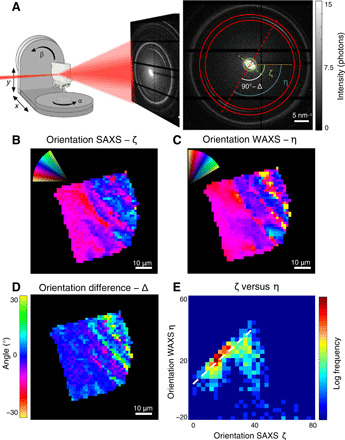
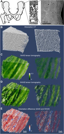
Bone is built from collagen fibrils and biomineral nanoparticles. In humans, they are organized in lamellar twisting patterns on the microscale. It has been a central tenet that the biomineral nanoparticles are co-aligned with the bone nanostructure. Here, we reconstruct the three-dimensional orientation in human lamellar bone of both the nanoscale features and the biomineral crystal lattice from small-angle x-ray scattering and wide-angle x-ray scattering, respectively. While most of the investigated regions show well-aligned nanostructure and crystal structure, consistent with current bone models, we report a localized difference in orientation distribution between the nanostructure and the biomineral crystals in specific bands. Our results show a robust and systematic, but localized, variation in the alignment of the two signals, which can be interpreted as either an additional mineral fraction in bone, a preferentially aligned extrafibrillar fraction, or the result of transverse stacking of mineral particles over several fibrils.
骨骼由胶原纤维和生物矿物纳米颗粒构成。在人类体内,它们在微观尺度上以层状扭曲模式排列。一直以来的核心观点是生物矿物纳米颗粒与骨纳米结构共同排列。在此,我们分别从小角X射线散射和广角X射线散射重建了人类层状骨中纳米级特征和生物矿物晶格的三维取向。虽然大多数研究区域显示出与当前骨模型一致的良好排列的纳米结构和晶体结构,但我们报告了特定条带中纳米结构和生物矿物晶体之间取向分布的局部差异。我们的结果显示了两种信号排列的稳健且系统但局部的变化,这可以解释为骨中额外的矿物部分、优先排列的纤维外部分,或者是矿物颗粒在多个纤维上横向堆叠的结果。