• 文献检索
  • 文档翻译
  • 深度研究
  • 学术资讯
  • Suppr Zotero 插件Zotero 插件
  • 邀请有礼
  • 套餐&价格
  • 历史记录
应用&插件
Suppr Zotero 插件Zotero 插件浏览器插件Mac 客户端Windows 客户端微信小程序
定价
高级版会员购买积分包购买API积分包
服务
文献检索文档翻译深度研究API 文档MCP 服务
关于我们
关于 Suppr公司介绍联系我们用户协议隐私条款
关注我们

Suppr 超能文献

核心技术专利:CN118964589B侵权必究
粤ICP备2023148730 号-1Suppr @ 2026

文献检索

告别复杂PubMed语法,用中文像聊天一样搜索,搜遍4000万医学文献。AI智能推荐,让科研检索更轻松。

立即免费搜索

文件翻译

保留排版,准确专业,支持PDF/Word/PPT等文件格式,支持 12+语言互译。

免费翻译文档

深度研究

AI帮你快速写综述,25分钟生成高质量综述,智能提取关键信息,辅助科研写作。

立即免费体验

冠状病毒感染的影像学表现:严重急性呼吸综合征冠状病毒、中东呼吸综合征冠状病毒和严重急性呼吸综合征冠状病毒 2。

Imaging findings in coronavirus infections: SARS-CoV, MERS-CoV, and SARS-CoV-2.

机构信息

Department of Radiology, Hospital de Sant Pau. Universidad Autónoma de Barcelona, Barcelona, Spain.

Department of Radiology and Biomedical Research Institute, Pusan National University Hospital, Busan, Korea.

出版信息

Br J Radiol. 2020 Aug;93(1112):20200515. doi: 10.1259/bjr.20200515. Epub 2020 Jul 6.

DOI:10.1259/bjr.20200515
PMID:32584604
原文链接:https://pmc.ncbi.nlm.nih.gov/articles/PMC7446008/
Abstract

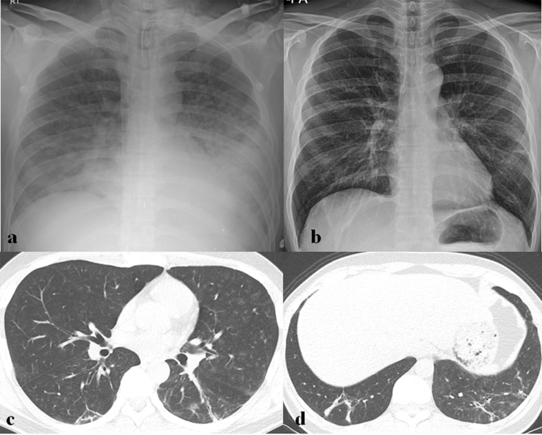
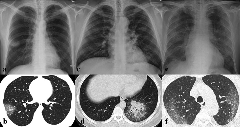
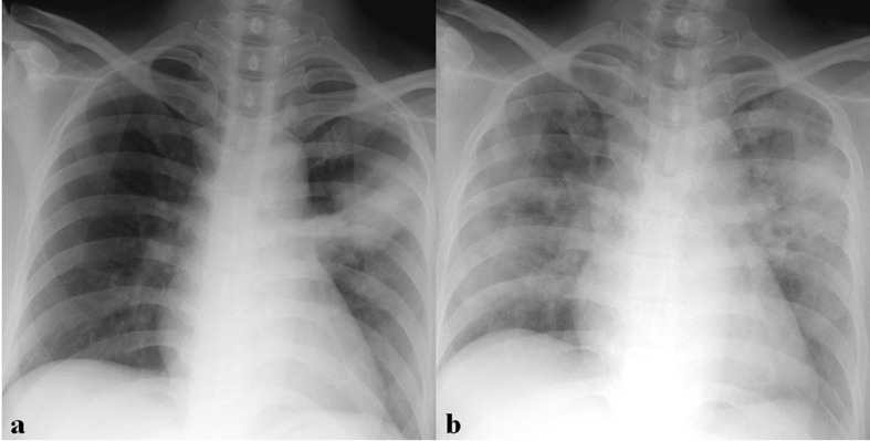
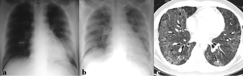
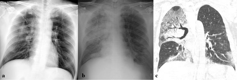
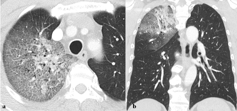
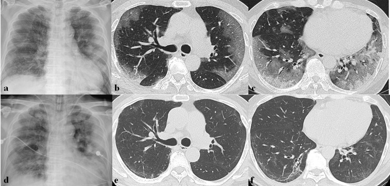
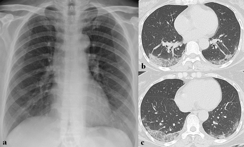
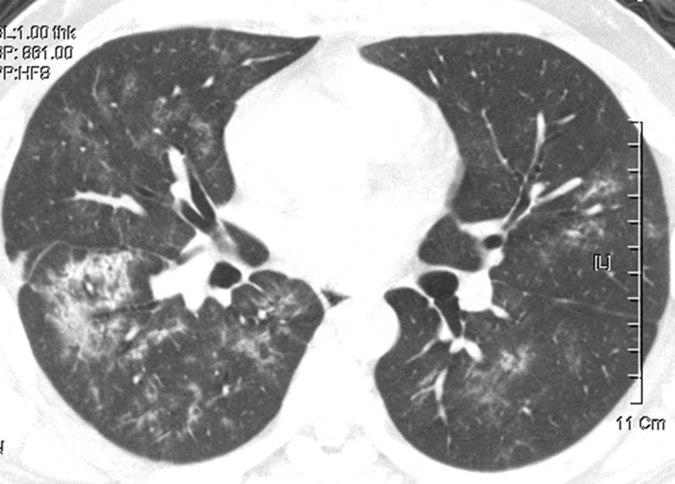
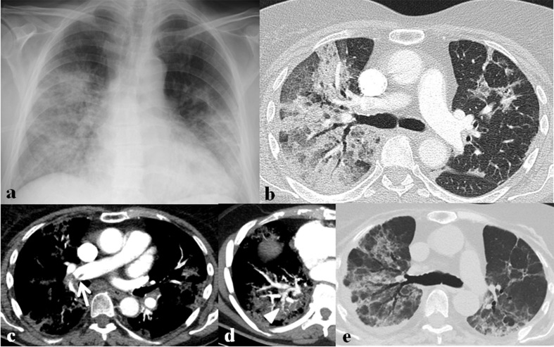
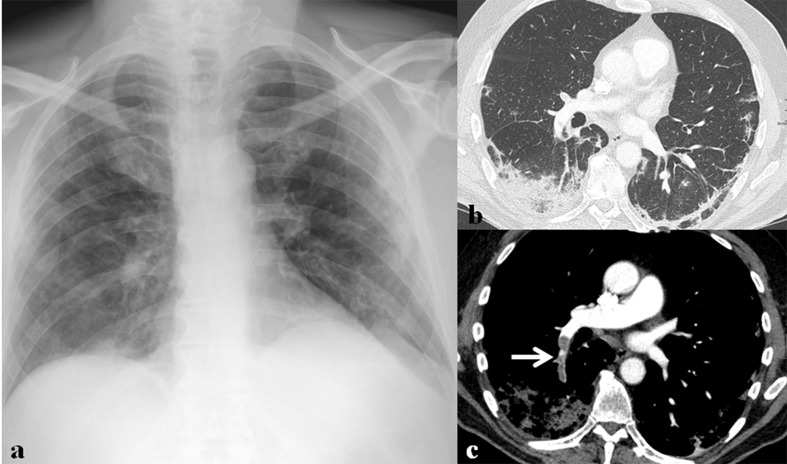
During the first two decades of the 21st century, there have been three coronavirus infection outbreaks raising global health concerns by severe acute respiratory syndrome coronavirus (SARS-CoV), the Middle East respiratory syndrome coronavirus (MERS-CoV), and the SARS-CoV-2. Although the reported imaging findings of coronavirus infection are variable and non-specific, the most common initial chest radiograph (CXR) and CT findings are ground-glass opacities and consolidation with peripheral predominance and eventually spread to involve both lungs as the disease progresses. These findings can be explained by the immune pathogenesis of coronavirus infection causing diffuse alveolar damage. Although it is insensitive in mild or early coronavirus infection, the CXR remains as the first-line and the most commonly used imaging modality. That is because it is rapid and easily accessible and helpful for monitoring patient progress during treatment. CT is more sensitive to detect early parenchymal lung abnormalities and disease progression, and can provide an alternative diagnosis. In this pictorial review, various coronavirus infection cases are presented to provide imaging spectrums of coronavirus infection and present differences in imaging among them or from other viral infections, and to discuss the role of imaging in viral infection outbreaks.

摘要

在 21 世纪的头二十年里,有三种冠状病毒感染爆发引起了全球健康关注,分别是严重急性呼吸综合征冠状病毒(SARS-CoV)、中东呼吸综合征冠状病毒(MERS-CoV)和 SARS-CoV-2。尽管冠状病毒感染的报告影像学表现多种多样且无特异性,但最常见的初始胸部 X 线摄影(CXR)和 CT 表现为磨玻璃影和实变影,以周边为主,随着疾病的进展逐渐累及双肺。这些发现可以用冠状病毒感染引起的免疫发病机制来解释,导致弥漫性肺泡损伤。虽然在轻度或早期冠状病毒感染中不敏感,但 CXR 仍然是一线和最常用的影像学检查方法。这是因为它快速、易于获得,有助于在治疗过程中监测患者的病情进展。CT 对检测早期肺实质异常和疾病进展更敏感,并能提供替代诊断。在这篇影像学综述中,展示了各种冠状病毒感染的病例,提供了冠状病毒感染的影像学谱,并介绍了它们之间或与其他病毒感染之间的影像学差异,并讨论了影像学在病毒感染爆发中的作用。

https://cdn.ncbi.nlm.nih.gov/pmc/blobs/cb66/7446008/ca6e6ad483f4/bjr.20200515.g015.jpg
https://cdn.ncbi.nlm.nih.gov/pmc/blobs/cb66/7446008/1cc52c64763a/bjr.20200515.g001.jpg
https://cdn.ncbi.nlm.nih.gov/pmc/blobs/cb66/7446008/49c6e802cdc8/bjr.20200515.g002.jpg
https://cdn.ncbi.nlm.nih.gov/pmc/blobs/cb66/7446008/e4fefbefd8e5/bjr.20200515.g003.jpg
https://cdn.ncbi.nlm.nih.gov/pmc/blobs/cb66/7446008/2755f1d266ff/bjr.20200515.g004.jpg
https://cdn.ncbi.nlm.nih.gov/pmc/blobs/cb66/7446008/baa343e9eec9/bjr.20200515.g005.jpg
https://cdn.ncbi.nlm.nih.gov/pmc/blobs/cb66/7446008/2b8317882c87/bjr.20200515.g006.jpg
https://cdn.ncbi.nlm.nih.gov/pmc/blobs/cb66/7446008/213b72357059/bjr.20200515.g007.jpg
https://cdn.ncbi.nlm.nih.gov/pmc/blobs/cb66/7446008/2cc7edd32743/bjr.20200515.g008.jpg
https://cdn.ncbi.nlm.nih.gov/pmc/blobs/cb66/7446008/3fd26731f025/bjr.20200515.g009.jpg
https://cdn.ncbi.nlm.nih.gov/pmc/blobs/cb66/7446008/582f22d836ab/bjr.20200515.g010.jpg
https://cdn.ncbi.nlm.nih.gov/pmc/blobs/cb66/7446008/27cf9e47df8b/bjr.20200515.g011.jpg
https://cdn.ncbi.nlm.nih.gov/pmc/blobs/cb66/7446008/a62db5d545af/bjr.20200515.g012.jpg
https://cdn.ncbi.nlm.nih.gov/pmc/blobs/cb66/7446008/4ea62e301590/bjr.20200515.g013.jpg
https://cdn.ncbi.nlm.nih.gov/pmc/blobs/cb66/7446008/3f5bdc44aa73/bjr.20200515.g014.jpg
https://cdn.ncbi.nlm.nih.gov/pmc/blobs/cb66/7446008/ca6e6ad483f4/bjr.20200515.g015.jpg

相似文献

1
Imaging findings in coronavirus infections: SARS-CoV, MERS-CoV, and SARS-CoV-2.冠状病毒感染的影像学表现:严重急性呼吸综合征冠状病毒、中东呼吸综合征冠状病毒和严重急性呼吸综合征冠状病毒 2。
Br J Radiol. 2020 Aug;93(1112):20200515. doi: 10.1259/bjr.20200515. Epub 2020 Jul 6.
2
Similarities and Differences of Early Pulmonary CT Features of Pneumonia Caused by SARS-CoV-2, SARS-CoV and MERS-CoV: Comparison Based on a Systemic Review.新型冠状病毒、严重急性呼吸综合征冠状病毒和中东呼吸综合征冠状病毒所致肺炎早期肺部CT特征的异同:基于系统评价的比较
Chin Med Sci J. 2020 Sep 30;35(3):254-261. doi: 10.24920/003727.
3
Pediatric SARS, H1N1, MERS, EVALI, and Now Coronavirus Disease (COVID-19) Pneumonia: What Radiologists Need to Know.儿童 SARS、H1N1、MERS、EVALI 及现在的新型冠状病毒病(COVID-19)肺炎:放射科医生需要了解的知识。
AJR Am J Roentgenol. 2020 Sep;215(3):736-744. doi: 10.2214/AJR.20.23267. Epub 2020 Apr 30.
4
Overview: The history and pediatric perspectives of severe acute respiratory syndromes: Novel or just like SARS.概述:严重急性呼吸系统综合征的历史和儿科视角:新型还是类似 SARS。
Pediatr Pulmonol. 2020 Jul;55(7):1584-1591. doi: 10.1002/ppul.24810. Epub 2020 Jun 1.
5
Imaging and clinical features of patients with 2019 novel coronavirus SARS-CoV-2.新型冠状病毒 2019 年 SARS-CoV-2 患者的影像学和临床特征。
Eur J Nucl Med Mol Imaging. 2020 May;47(5):1275-1280. doi: 10.1007/s00259-020-04735-9. Epub 2020 Feb 28.
6
Clinical characteristics of COVID-19 in 104 people with SARS-CoV-2 infection on the Diamond Princess cruise ship: a retrospective analysis.《104 名“钻石公主”号邮轮上 SARS-CoV-2 感染患者的 COVID-19 临床特征:一项回顾性分析》。
Lancet Infect Dis. 2020 Sep;20(9):1043-1050. doi: 10.1016/S1473-3099(20)30482-5. Epub 2020 Jun 12.
7
Molecular Basis of Pathogenesis of Coronaviruses: A Comparative Genomics Approach to Planetary Health to Prevent Zoonotic Outbreaks in the 21st Century.冠状病毒发病机制的分子基础:以比较基因组学方法研究行星健康,以预防 21 世纪的人畜共患病爆发。
OMICS. 2020 Nov;24(11):634-644. doi: 10.1089/omi.2020.0131. Epub 2020 Sep 16.
8
Coronavirus infections in children: from SARS and MERS to COVID-19, a narrative review of epidemiological and clinical features.儿童冠状病毒感染:从SARS、MERS到COVID-19,关于流行病学和临床特征的叙述性综述
Acta Biomed. 2020 Sep 7;91(3):e2020032. doi: 10.23750/abm.v91i3.10294.
9
Diagnostic value and key features of computed tomography in Coronavirus Disease 2019.计算机断层扫描在 2019 年冠状病毒病中的诊断价值和关键特征。
Emerg Microbes Infect. 2020 Dec;9(1):787-793. doi: 10.1080/22221751.2020.1750307.
10
Diagnosis of SARS-CoV-2 infection based on CT scan vs RT-PCR: reflecting on experience from MERS-CoV.基于CT扫描与逆转录聚合酶链反应的新型冠状病毒2感染诊断:对中东呼吸综合征冠状病毒经验的反思
J Hosp Infect. 2020 Jun;105(2):154-155. doi: 10.1016/j.jhin.2020.03.001. Epub 2020 Mar 6.

引用本文的文献

1
Short- and Long-Term Chest-CT Findings after Recovery from COVID-19: A Systematic Review and Meta-Analysis.新冠康复后的短期和长期胸部CT表现:一项系统评价和荟萃分析
Diagnostics (Basel). 2024 Mar 14;14(6):621. doi: 10.3390/diagnostics14060621.
2
Comparative imaging findings among different primary betacoronaviruses.不同原发性β冠状病毒之间的对比影像学表现。
Pol J Radiol. 2022 Nov 30;87:e641-e651. doi: 10.5114/pjr.2022.121763. eCollection 2022.
3
Vaccine effectiveness against referral to hospital after SARS-CoV-2 infection in St. Petersburg, Russia, during the Delta variant surge: a test-negative case-control study.

本文引用的文献

1
Segmental Pulmonary Vascular Changes in COVID-19 Pneumonia.新型冠状病毒肺炎的节段性肺血管改变
AJR Am J Roentgenol. 2020 Sep;215(3):W33. doi: 10.2214/AJR.20.23443. Epub 2020 May 8.
2
Incidence of thrombotic complications in critically ill ICU patients with COVID-19.COVID-19 重症监护病房危重症患者的血栓并发症发生率。
Thromb Res. 2020 Jul;191:145-147. doi: 10.1016/j.thromres.2020.04.013. Epub 2020 Apr 10.
3
Molecular immune pathogenesis and diagnosis of COVID-19.新型冠状病毒肺炎的分子免疫发病机制与诊断
俄罗斯圣彼得堡在德尔塔变异株流行期间,针对感染 SARS-CoV-2 后转诊至医院的疫苗效力:一项病例对照研究。
BMC Med. 2022 Sep 20;20(1):312. doi: 10.1186/s12916-022-02509-8.
4
Usefulness of Hospital Admission Chest X-ray Score for Predicting Mortality and ICU Admission in COVID-19 Patients.医院入院胸部X线评分对预测COVID-19患者死亡率和入住重症监护病房的价值
J Clin Med. 2022 Jun 20;11(12):3548. doi: 10.3390/jcm11123548.
5
An Imaging Overview of COVID-19 ARDS in ICU Patients and Its Complications: A Pictorial Review.ICU 患者中 COVID-19 急性呼吸窘迫综合征及其并发症的影像学概述:图文综述
Diagnostics (Basel). 2022 Mar 29;12(4):846. doi: 10.3390/diagnostics12040846.
6
Clinical characteristics of patients with confirmed and suspected COVID-19.确诊和疑似新冠肺炎患者的临床特征
Am J Transl Res. 2021 Nov 15;13(11):12897-12904. eCollection 2021.
7
Update Advances on C-Reactive Protein in COVID-19 and Other Viral Infections.更新:C 反应蛋白在 COVID-19 及其他病毒感染中的研究进展。
Front Immunol. 2021 Aug 10;12:720363. doi: 10.3389/fimmu.2021.720363. eCollection 2021.
8
Current diagnostic approaches to detect two important betacoronaviruses: Middle East respiratory syndrome coronavirus (MERS-CoV) and severe acute respiratory syndrome coronavirus 2 (SARS-CoV-2).目前用于检测两种重要的贝塔冠状病毒的诊断方法:中东呼吸综合征冠状病毒(MERS-CoV)和严重急性呼吸综合征冠状病毒 2(SARS-CoV-2)。
Pathol Res Pract. 2021 Sep;225:153565. doi: 10.1016/j.prp.2021.153565. Epub 2021 Jul 24.
9
Lung donation following SARS-CoV-2 infection.新冠病毒感染后的肺脏捐献。
Am J Transplant. 2021 Dec;21(12):4073-4078. doi: 10.1111/ajt.16777. Epub 2021 Aug 9.
10
Correlation between Chest X-Ray Severity in COVID-19 and Age in Mexican-Mestizo Patients: An Observational Cross-Sectional Study.墨西哥梅斯蒂索患者中 COVID-19 的胸部 X 光严重程度与年龄的相关性:一项观察性横断面研究。
Biomed Res Int. 2021 Apr 29;2021:5571144. doi: 10.1155/2021/5571144. eCollection 2021.
J Pharm Anal. 2020 Apr;10(2):102-108. doi: 10.1016/j.jpha.2020.03.001. Epub 2020 Mar 5.
4
The Role of Chest Imaging in Patient Management during the COVID-19 Pandemic: A Multinational Consensus Statement from the Fleischner Society.《COVID-19 大流行期间患者管理中的胸部影像学作用:Fleischner 学会的一项多国共识声明》。
Radiology. 2020 Jul;296(1):172-180. doi: 10.1148/radiol.2020201365. Epub 2020 Apr 7.
5
Frequency and Distribution of Chest Radiographic Findings in Patients Positive for COVID-19.COVID-19 阳性患者的胸部 X 线表现的频率和分布。
Radiology. 2020 Aug;296(2):E72-E78. doi: 10.1148/radiol.2020201160. Epub 2020 Mar 27.
6
Temporal Changes of CT Findings in 90 Patients with COVID-19 Pneumonia: A Longitudinal Study.90 例 COVID-19 肺炎患者 CT 表现的时间变化:一项纵向研究。
Radiology. 2020 Aug;296(2):E55-E64. doi: 10.1148/radiol.2020200843. Epub 2020 Mar 19.
7
Risk Factors Associated With Acute Respiratory Distress Syndrome and Death in Patients With Coronavirus Disease 2019 Pneumonia in Wuhan, China.中国武汉 2019 年冠状病毒病肺炎患者急性呼吸窘迫综合征和死亡的相关危险因素。
JAMA Intern Med. 2020 Jul 1;180(7):934-943. doi: 10.1001/jamainternmed.2020.0994.
8
CT Imaging Features of 2019 Novel Coronavirus (2019-nCoV).2019 新型冠状病毒(2019-nCoV)的 CT 影像学特征。
Radiology. 2020 Apr;295(1):202-207. doi: 10.1148/radiol.2020200230. Epub 2020 Feb 4.
9
Clinical implication of radiographic scores in acute Middle East respiratory syndrome coronavirus pneumonia: Report from a single tertiary-referral center of South Korea.急性中东呼吸综合征冠状病毒肺炎影像学评分的临床意义:来自韩国一家三级转诊中心的报告。
Eur J Radiol. 2018 Oct;107:196-202. doi: 10.1016/j.ejrad.2018.09.008. Epub 2018 Sep 11.
10
Radiographic and CT Features of Viral Pneumonia.病毒性肺炎的影像学和 CT 特征。
Radiographics. 2018 May-Jun;38(3):719-739. doi: 10.1148/rg.2018170048.