Department of Rheumatology & Clinical Immunology, University Medical Centre Utrecht, Utrecht, The Netherlands.
Department of Anatomy, Histology and Embryology, Faculty of Medicine, University of Debrecen, Debrecen, Hungary.
BMC Mol Cell Biol. 2020 Jun 26;21(1):47. doi: 10.1186/s12860-020-00288-9.
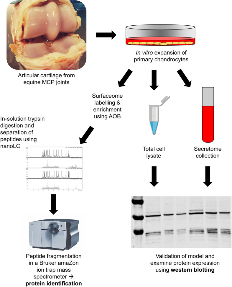
Chondrocytes are exposed to an inflammatory micro-environment in the extracellular matrix (ECM) of articular cartilage in joint diseases such as osteoarthritis (OA) and rheumatoid arthritis (RA). In OA, degenerative changes and low-grade inflammation within the joint transform the behaviour and metabolism of chondrocytes, disturb the balance between ECM synthesis and degradation, and alter the osmolality and ionic composition of the micro-environment. We hypothesize that chondrocytes adjust their physiology to the inflammatory microenvironment by modulating the expression of cell surface proteins, collectively referred to as the 'surfaceome'. Therefore, the aim of this study was to characterize the surfaceome of primary equine chondrocytes isolated from healthy joints following exposure to the pro-inflammatory cytokines interleukin-1-beta (IL-1β) and tumour necrosis factor-alpha (TNF-α). We employed combined methodology that we recently developed for investigating the surfaceome in stem cells. Membrane proteins were isolated using an aminooxy-biotinylation technique and analysed by mass spectrometry using high throughput shotgun proteomics. Selected proteins were validated by western blotting.
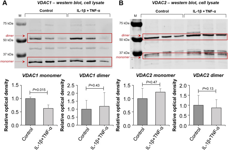
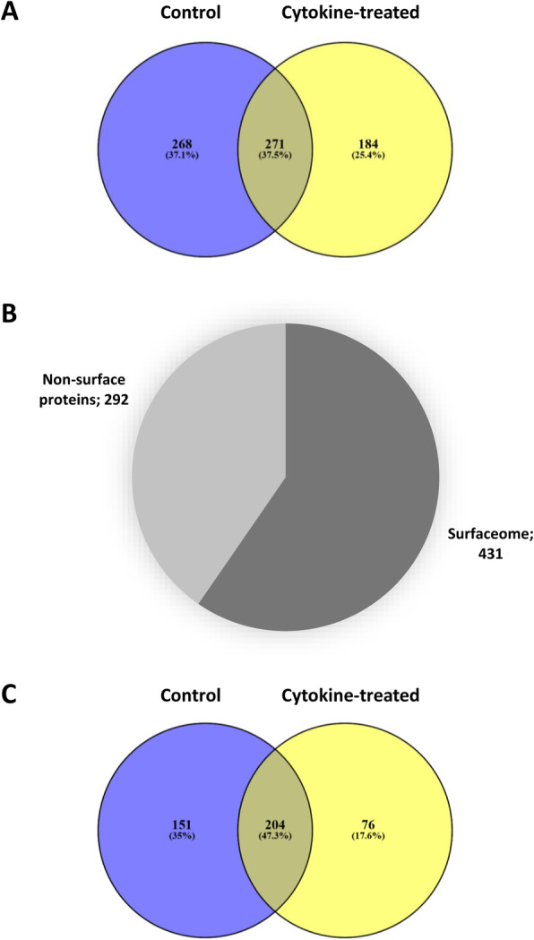
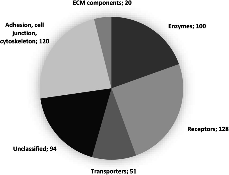
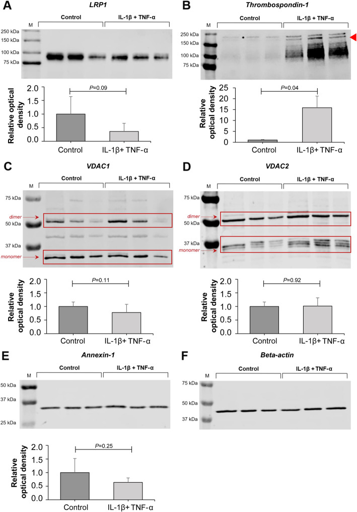
Amongst the 431 unique cell surface proteins identified, a high percentage of low-abundance proteins, such as ion channels, receptors and transporter molecules were detected. Data are available via ProteomeXchange with identifier PXD014773. A high number of proteins exhibited different expression patterns following chondrocyte stimulation with pro-inflammatory cytokines. Low density lipoprotein related protein 1 (LPR-1), thrombospondin-1 (TSP-1), voltage dependent anion channel (VDAC) 1-2 and annexin A1 were considered to be of special interest and were analysed further by western blotting.
Our results provide, for the first time, a repository for proteomic data on differentially expressed low-abundance membrane proteins on the surface of chondrocytes in response to pro-inflammatory stimuli.
软骨细胞在骨关节炎(OA)和类风湿关节炎(RA)等关节疾病的关节软骨细胞外基质(ECM)中会受到炎症微环境的影响。在 OA 中,关节内的退行性变化和低水平炎症会改变软骨细胞的行为和代谢,扰乱 ECM 合成与降解之间的平衡,并改变微环境的渗透压和离子组成。我们假设软骨细胞通过调节细胞表面蛋白的表达来调节其生理机能,这些蛋白统称为“表面组”。因此,本研究的目的是在从健康关节中分离的原代马软骨细胞暴露于促炎细胞因子白细胞介素 1-β(IL-1β)和肿瘤坏死因子-α(TNF-α)后,对其表面组进行特征描述。我们采用了我们最近开发的用于研究干细胞表面组的组合方法。使用氨氧基生物素化技术分离膜蛋白,并使用高通量 shotgun 蛋白质组学进行质谱分析。通过 Western blot 验证选择的蛋白质。
在鉴定的 431 个独特的细胞表面蛋白中,检测到了高比例的低丰度蛋白,如离子通道、受体和转运体分子。数据可通过 ProteomeXchange 以标识符 PXD014773 获得。在软骨细胞受到促炎细胞因子刺激后,大量蛋白质表现出不同的表达模式。低密度脂蛋白相关蛋白 1(LPR-1)、血小板反应蛋白-1(TSP-1)、电压依赖性阴离子通道(VDAC)1-2 和膜联蛋白 A1 被认为特别有趣,并通过 Western blot 进一步分析。
我们的结果首次提供了一个储存库,用于存储在受到促炎刺激后,软骨细胞表面差异表达的低丰度膜蛋白的蛋白质组学数据。