Ma Ziwei, Pan Xuefeng, Zhou Danni, Zhu Zhuangzhi, Xu Aiping, Shi Peng, Chen Hong
Department of Ophthalmology, the First People's Hospital of Huzhou, Huzhou, Zhejiang Province, China.
Medicine (Baltimore). 2020 Jun 26;99(26):e21007. doi: 10.1097/MD.0000000000021007.
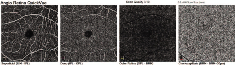
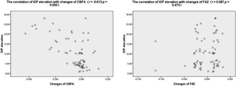
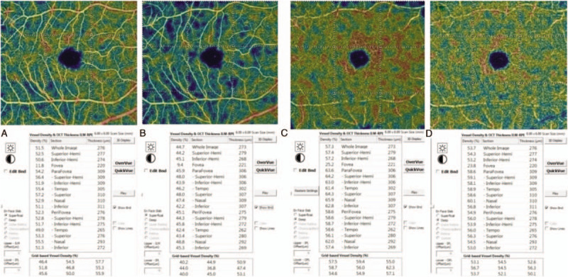
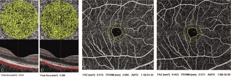
The aim of this study was to investigate the changes of retinal vessel density (VD) and choriocapillary blood flow area (CBFA) in macula after an acute intraocular pressure (IOP) elevation observed using optical coherence tomography angiography.This was a prospective comparative study of subjects with narrow anterior chamber angles who underwent laser peripheral iridotomies (LPIs). The IOP was measured before and 1 hour after the LPI. The retinal VDs and CBFAs of the macula were measured using optical coherence tomography angiography at the baseline and 1 hour after the LPI.A total of 88 eyes of 88 individuals were enrolled in our study, and 70 eyes of 70 individuals finally completed the study with a mean IOP rise of 10.2 ± 7.5 mm Hg after the LPI. The VDs and areas of foveal avascular zone of all of the subjects did not differ significantly between the measurements obtained at the baseline and 1 hour after the LPI (P > .05). However, there were statistically significant differences in the CBFAs at the baseline and 1 hour after the LPI (P < .05). Based on the magnitude of the rise in the IOP, we divided the subjects into three groups: group A = IOP rise ≤ 10 mm Hg, group B = 10 mm Hg < IOP rise ≤20 mm Hg, and group C = IOP rise > 20 mmHg. The VDs of the macula measured at the baseline were significantly different from the measurements obtained 1 hour after the LPI in group C in either the superficial retinal layer or deep retinal layer (P < .05). Compared with baseline, the CBFAs measured at 1 hour after the LPI were decreased in group B and group C (P < .05).In these subjects with narrow antenior chamber, the blood flow in macula began to be affected with the acute IOP rise greater than 10 mm Hg. It was confirmed that the retina and choroid showed some different ability to regulate its blood flow in response to changes in IOP.
本研究旨在利用光学相干断层扫描血管造影术,观察急性眼压升高后黄斑区视网膜血管密度(VD)和脉络膜毛细血管血流面积(CBFA)的变化。这是一项对接受激光周边虹膜切开术(LPI)的窄前房角受试者进行的前瞻性对照研究。在LPI术前和术后1小时测量眼压。使用光学相干断层扫描血管造影术在基线和LPI术后1小时测量黄斑区的视网膜VD和CBFA。
本研究共纳入88例个体的88只眼,最终70例个体的70只眼完成研究,LPI术后平均眼压升高10.2±7.5 mmHg。所有受试者在基线和LPI术后1小时测量的黄斑区VD和无血管区面积差异无统计学意义(P>0.05)。然而,基线和LPI术后1小时的CBFA差异有统计学意义(P<0.05)。根据眼压升高幅度,将受试者分为三组:A组=眼压升高≤10 mmHg,B组=10 mmHg<眼压升高≤20 mmHg,C组=眼压升高>20 mmHg。C组在浅层视网膜层或深层视网膜层,基线时测量的黄斑区VD与LPI术后1小时测量值相比差异有统计学意义(P<0.05)。与基线相比,B组和C组在LPI术后1小时测量的CBFA降低(P<0.05)。
在这些窄前房受试者中,急性眼压升高大于10 mmHg时,黄斑区血流开始受到影响。证实视网膜和脉络膜在应对眼压变化时调节血流的能力有所不同。