Thoracic Surgery Department, Istituto di Ricovero e Cura a Carattere Scientifico MultiMedica, Milan, Italy.
Oncology and Hemato-Oncology Department, University of Milan, and Inter-Hospital Pathology Division, Istituto di Ricovero e Cura a Carattere Scientifico MultiMedica, Milan, Italy.
Ann Thorac Surg. 2021 Jan;111(1):e23-e25. doi: 10.1016/j.athoracsur.2020.06.008. Epub 2020 Jun 27.
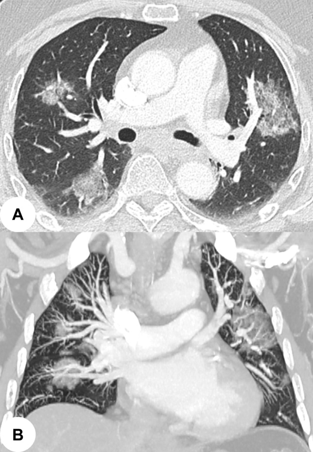
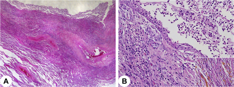
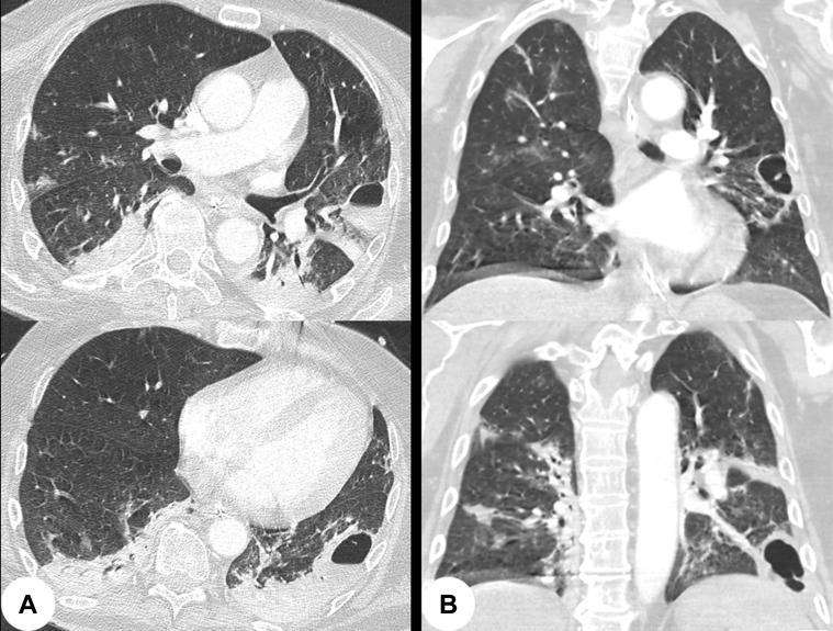
Emerging studies on radiologic findings in patients with coronavirus disease 2019 (COVID-19) report a high incidence of bilateral lung involvement, with ground-glass opacities imaging being the most common pattern on computed tomography. Cystic lesions, such as pneumatoceles, are rare, although they may occur in 10% of cases. Cyst formation may be explained by a focal pulmonary trauma caused by mechanical ventilation or infection-related damage to the alveolar walls leading to pneumatoceles. The superinfection of pneumatoceles is a potential life-threatening condition for which no standardized therapeutic algorithm has been accepted. We report a case of a COVID-19 patient successfully treated by lung resections for infected pneumatoceles.
关于 2019 冠状病毒病(COVID-19)患者的放射学表现的新兴研究报告称,双肺受累的发生率很高,计算机断层扫描(CT)最常见的表现为磨玻璃影。尽管囊腔(如肺大疱)在 10%的病例中可能发生,但较为罕见。囊腔的形成可能是由于机械通气引起的局灶性肺损伤或与感染相关的肺泡壁损伤导致肺大疱。肺大疱的继发感染是一种潜在的危及生命的情况,但目前尚未接受任何标准化的治疗方案。我们报告了一例 COVID-19 患者,该患者因感染性肺大疱而成功接受了肺切除术治疗。