Hubei Key Laboratory of Embryonic Stem Cell Research, Taihe Hospital, Hubei University of Medicine, Shiyan, 442000, Hubei, China.
Cardiovascular Department, Hubei University of Medicine, Taihe Hospital, Hubei University of Medicine, Shiyan, 442000, Hubei, China.
BMC Complement Med Ther. 2020 Jul 2;20(1):203. doi: 10.1186/s12906-020-02992-7.
Qiliqiangxin (QLQX) is a preparation refined from a traditional Chinese medicine compound. It plays an important role in protecting cardiac function after myocardial infarction (MI). However, the underline mechanism of QLQX action is not clear. The purpose of this study was to detect the effects of QLQX on mitophagy after MI.
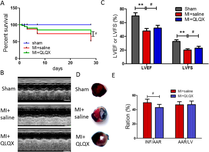
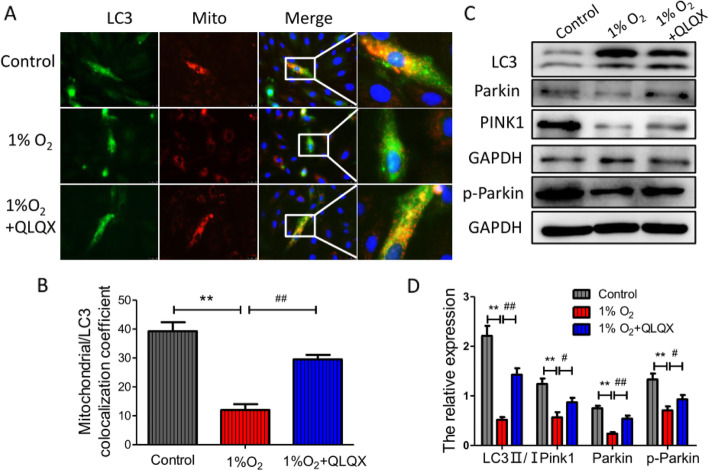
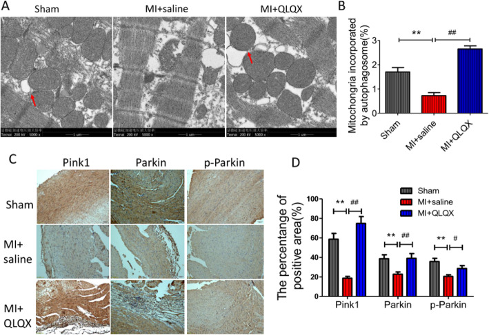
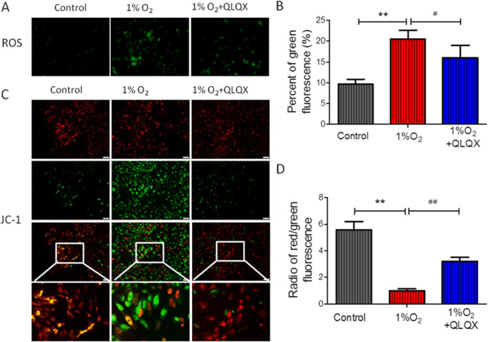
Male FVB/NJ mice aged 8-10 weeks were underwent left coronary artery ligation and were orally administered either QLQX (0.25 g/kg/d) or saline. Twenty-eight days after surgical operation, the cardiac function of mice was detected by echocardiography. Electron Microscopy was used to observe the microstructure of cardiomyocytes. Myocardial apoptosis was examined by TdT-mediated dUTP Nick-End Labeling (TUNEL) and western blot. H9c2 cells were cultured in a hypoxic incubator chamber (5% CO, 1% O, 94% N) for 12 h and pretreated with or without QLQX (0.5 mg/mL). The cell apoptosis, reactive oxygen species (ROS), mitochondrial membrane potential and mitophagy were detected.
When compared to sham group, the cardiac function of MI mice decreased significantly, and their cardiomyocyte apoptosis and mitochondrial damage were more serious. These MI-induced cardiac changes could be reversed by QLQX treatment. In vitro experiments also confirmed that QLQX could protect cardiomyocytes from hypoxia-induced apoptosis and mitochondrial damage. Further study indicated that QLQX could increase the expression of Pink1 and Parkin in cardiomyocytes.
Qiliqiangxin could reduce cardiomyocytes apotosis and improved heart function in infarcted heart through Pink1-mediated mitochondrial autophagy.
芪苈强心是一种从中药复方中精制而成的制剂。它在心肌梗死后保护心脏功能方面发挥着重要作用。然而,芪苈强心的作用机制尚不清楚。本研究旨在检测芪苈强心对心肌梗死后自噬的影响。
雄性 FVB/NJ 小鼠 8-10 周龄,行左冠状动脉结扎术,给予芪苈强心(0.25 g/kg/d)或生理盐水口服。手术后 28 天,通过超声心动图检测小鼠心功能。电子显微镜观察心肌细胞的微观结构。通过 TdT 介导的 dUTP 缺口末端标记法(TUNEL)和蛋白质印迹法检测心肌细胞凋亡。将 H9c2 细胞置于低氧孵育箱(5% CO、1% O、94% N)中培养 12 h,并用或不用芪苈强心(0.5 mg/mL)预处理。检测细胞凋亡、活性氧(ROS)、线粒体膜电位和自噬。
与假手术组相比,心肌梗死小鼠的心脏功能明显下降,心肌细胞凋亡和线粒体损伤更为严重。芪苈强心治疗可逆转这些 MI 引起的心脏变化。体外实验也证实,芪苈强心可以保护心肌细胞免受低氧诱导的凋亡和线粒体损伤。进一步的研究表明,芪苈强心可以增加心肌细胞中 Pink1 和 Parkin 的表达。
芪苈强心通过 Pink1 介导的线粒体自噬减少心肌细胞凋亡,改善梗死心脏的心功能。