Gao Wenwen, Han Xiaowei, Li Haimei, Zhu Yijiang, Du Lei, Wang Yuli, Shi Sumin, Liu Jing, Fu Chao, Zhang Lu, Ma Guolin
Department of Radiology, China-Japan Friendship Hospital, Beijing, China.
Department of Radiology, Fu Xing Hospital, Capital Medical University, Beijing, China.
Ann Transl Med. 2020 Jun;8(11):699. doi: 10.21037/atm.2020.03.133.
Dysarthria is one of the common symptoms of facial paralysis (FP). This study aimed to investigate functional alterations in the brain language network in early idiopathic peripheral FP patients with dysarthria using resting-state functional magnetic resonance imaging (fMRI).
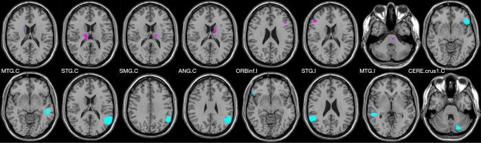
A total of 45 cases of FP (left 22, right 23) and 34 cases of healthy control (HC) were recruited into this study. The data of patients with left-side FP and matched controls (17 cases) were flipped from left to right, and the brain regions were defined as ipsilateral and contralateral regions. The FC of 16 ROIs in classical language centers and regions that may be involved in language function were calculated. After identifying the differences of FC between the two groups, the correlation analysis between altered FC and TFGS score of oral muscle movement in FP group were analyzed.
The FC between bilateral language regions has a significantly decreased trend in FP group compared with HC group (P<0.05). The ipsilateral inferior frontal gyrus, superior temporal gyrus, and middle temporal gyrus exhibited significantly decreased FC with multiple brain regions. In addition, we found that thalamus and cerebellum also with a significant alteration in FC in FP patients indicating that these two regions may also be involved in the mechanism of dysarthria in FP. The correlation analysis results indicated that the decrease of FC was positively correlated with the severity of oral paralysis.
Idiopathic peripheral FP with dysarthria induces several FC alterations in the brain language network. The severity of oral paralysis is associated with these functional alterations.
构音障碍是面瘫(FP)的常见症状之一。本研究旨在使用静息态功能磁共振成像(fMRI)研究早期特发性周围性面瘫伴构音障碍患者脑语言网络的功能改变。
本研究共纳入45例面瘫患者(左侧22例,右侧23例)和34例健康对照(HC)。将左侧面瘫患者及匹配对照(17例)的数据左右翻转,并将脑区定义为同侧和对侧区域。计算经典语言中枢及可能参与语言功能的区域中16个感兴趣区(ROI)的功能连接(FC)。在确定两组之间FC的差异后,分析FP组中FC改变与口腔肌肉运动TFGS评分之间的相关性分析。
与HC组相比,FP组双侧语言区域之间的FC有显著下降趋势(P<0.05)。同侧额下回、颞上回和颞中回与多个脑区的FC显著降低。此外,我们发现丘脑和小脑在FP患者中FC也有显著改变,表明这两个区域可能也参与了FP构音障碍的机制。相关性分析结果表明,FC的降低与口腔麻痹的严重程度呈正相关。
特发性周围性面瘫伴构音障碍会导致脑语言网络中的多个FC改变。口腔麻痹的严重程度与这些功能改变有关。