• 文献检索
  • 文档翻译
  • 深度研究
  • 学术资讯
  • Suppr Zotero 插件Zotero 插件
  • 邀请有礼
  • 套餐&价格
  • 历史记录
应用&插件
Suppr Zotero 插件Zotero 插件浏览器插件Mac 客户端Windows 客户端微信小程序
定价
高级版会员购买积分包购买API积分包
服务
文献检索文档翻译深度研究API 文档MCP 服务
关于我们
关于 Suppr公司介绍联系我们用户协议隐私条款
关注我们

Suppr 超能文献

核心技术专利:CN118964589B侵权必究
粤ICP备2023148730 号-1Suppr @ 2026

文献检索

告别复杂PubMed语法,用中文像聊天一样搜索,搜遍4000万医学文献。AI智能推荐,让科研检索更轻松。

立即免费搜索

文件翻译

保留排版,准确专业,支持PDF/Word/PPT等文件格式,支持 12+语言互译。

免费翻译文档

深度研究

AI帮你快速写综述,25分钟生成高质量综述,智能提取关键信息,辅助科研写作。

立即免费体验

运用多模态成像描绘解剖结构与功能:巨大右冠状动脉瘤病例报告

Delineating anatomy and function with multimodality imaging: a case report of giant right coronary artery aneurysms.

作者信息

Kassab Kameel, Iskander Mina, Malhotra Saurabh, Pyslar Nataliya

机构信息

Division of Cardiology, Department of Cardiology, John H. Stroger, Jr. Hospital of Cook County, 1901 W. Harrison Street, Room 3620, Chicago, IL 60612, USA.

出版信息

Eur Heart J Case Rep. 2020 Apr 24;4(3):1-5. doi: 10.1093/ehjcr/ytaa089. eCollection 2020 Jun.

DOI:10.1093/ehjcr/ytaa089
PMID:32617482
原文链接:https://pmc.ncbi.nlm.nih.gov/articles/PMC7319827/
Abstract

BACKGROUND

Giant coronary artery aneurysms (CAAs) are rare and have been reported in patients with connective tissue diseases, arteritides, and atherosclerosis. Given the rarity of the condition, multimodality imaging is essential for comprehensive evaluation of coronary aneurysms and determination of their haemodynamic significance.

CASE SUMMARY

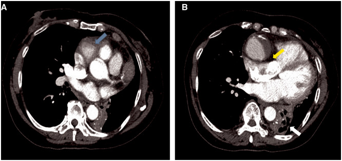
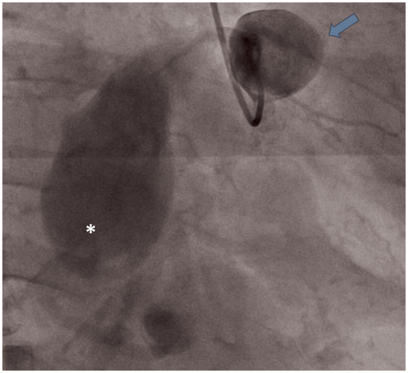
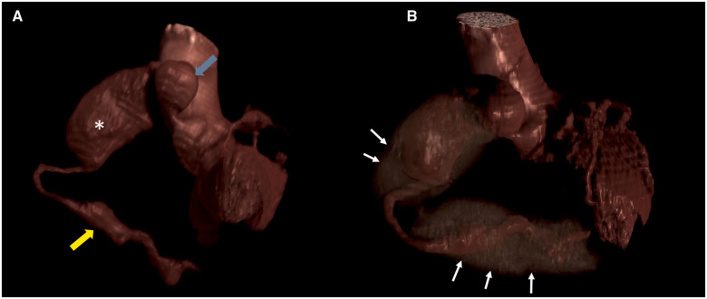
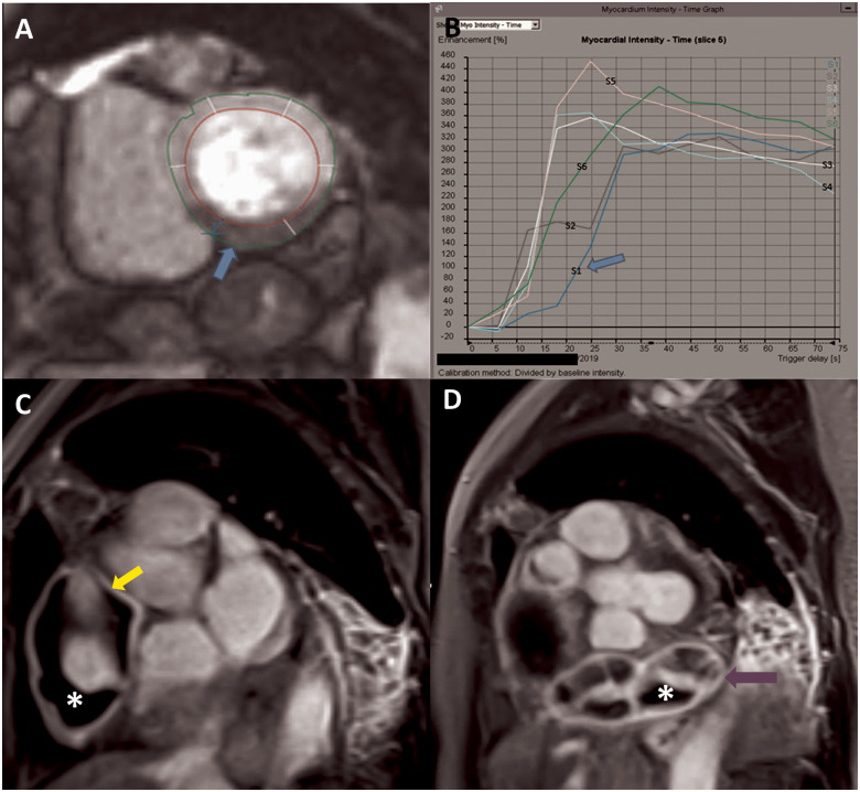
A 58-year-old Filipino female was evaluated for dyspnoea on exertion of one month. Chest computed tomography (CT) showed right coronary artery (RCA) aneurysms. Invasive coronary angiogram (ICA) confirmed two giant aneurysms of the RCA. Distal RCA could not be opacified due to contrast stagnation in the proximal aneurysms. Coronary CT angiography (CCTA) depicted an additional giant distal RCA aneurysm not visualized on ICA with intraluminal thrombosis. Contrast-enhanced cardiac magnetic resonance imaging (CMR) revealed delayed time to peak perfusion in the mid to apical inferior walls, on first-pass imaging, without myocardial scarring. Late gadolinium images revealed aneurysmal wall inflammation.

DISCUSSION

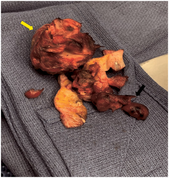
This case highlights the anatomical findings of giant CAA and the application of multimodality imaging for their accurate characterization. While ICA confirmed the presence of the aneurysms, CCTA enabled the assessment of their full extent and depict intraluminal thrombosis. Contrast-enhanced CMR delineated aneurysm wall characteristics, with first-pass images demonstrating reduced inferior wall perfusion at rest, which was likely the cause of patient's exertional symptoms. Management of giant coronary aneurysms involves surgical resection with bypass grafting.

摘要

背景

巨大冠状动脉瘤(CAA)较为罕见,已在结缔组织病、动脉炎和动脉粥样硬化患者中报道。鉴于该病症的罕见性,多模态成像对于冠状动脉瘤的全面评估及其血流动力学意义的确定至关重要。

病例摘要

一名58岁的菲律宾女性因劳力性呼吸困难1个月接受评估。胸部计算机断层扫描(CT)显示右冠状动脉(RCA)瘤。有创冠状动脉造影(ICA)证实RCA有两个巨大动脉瘤。由于近端动脉瘤内造影剂滞留,RCA远端未显影。冠状动脉CT血管造影(CCTA)显示一个在ICA上未显示的额外巨大RCA远端动脉瘤,并伴有腔内血栓形成。对比增强心脏磁共振成像(CMR)显示首次通过成像时,中至心尖下壁的峰值灌注延迟,无心肌瘢痕。延迟钆增强图像显示动脉瘤壁炎症。

讨论

本病例突出了巨大CAA的解剖学发现以及多模态成像在其准确特征描述中的应用。虽然ICA证实了动脉瘤的存在,但CCTA能够评估其全貌并显示腔内血栓形成。对比增强CMR描绘了动脉瘤壁特征,首次通过图像显示静息时下壁灌注减少,这可能是患者劳力性症状的原因。巨大冠状动脉瘤的治疗包括手术切除并进行搭桥移植。

https://cdn.ncbi.nlm.nih.gov/pmc/blobs/74fa/7319827/b9d7b6e40949/ytaa089f7.jpg
https://cdn.ncbi.nlm.nih.gov/pmc/blobs/74fa/7319827/c61ab9f57c17/ytaa089f1.jpg
https://cdn.ncbi.nlm.nih.gov/pmc/blobs/74fa/7319827/ec257f9b4286/ytaa089f2.jpg
https://cdn.ncbi.nlm.nih.gov/pmc/blobs/74fa/7319827/180e309d87a4/ytaa089f3.jpg
https://cdn.ncbi.nlm.nih.gov/pmc/blobs/74fa/7319827/3dc201b6021f/ytaa089f4.jpg
https://cdn.ncbi.nlm.nih.gov/pmc/blobs/74fa/7319827/d94be77c3701/ytaa089f5.jpg
https://cdn.ncbi.nlm.nih.gov/pmc/blobs/74fa/7319827/83f54b53e385/ytaa089f6.jpg
https://cdn.ncbi.nlm.nih.gov/pmc/blobs/74fa/7319827/b9d7b6e40949/ytaa089f7.jpg

相似文献

1
Delineating anatomy and function with multimodality imaging: a case report of giant right coronary artery aneurysms.运用多模态成像描绘解剖结构与功能:巨大右冠状动脉瘤病例报告
Eur Heart J Case Rep. 2020 Apr 24;4(3):1-5. doi: 10.1093/ehjcr/ytaa089. eCollection 2020 Jun.
2
Usefulness of multimodality cardiac imaging in a patient with ST elevation myocardial infarction caused by two giant coronary artery aneurysms.多模态心脏成像在一名由两个巨大冠状动脉瘤引起的ST段抬高型心肌梗死患者中的应用价值
BMJ Case Rep. 2019 Aug 4;12(8):e229995. doi: 10.1136/bcr-2019-229995.
3
Multiple giant coronary artery aneurysms combined with right coronary artery-pulmonary artery fistula: a case report.多发性巨大冠状动脉瘤合并右冠状动脉-肺动脉瘘:一例报告
BMC Surg. 2019 Jul 5;19(1):80. doi: 10.1186/s12893-019-0547-z.
4
Giant Coronary Aneurysm Causing Ostial Occlusion of Coronary Artery by Mass Effect: A Case Report.巨大冠状动脉瘤通过占位效应导致冠状动脉开口闭塞:一例报告
Cureus. 2021 Jul 9;13(7):e16280. doi: 10.7759/cureus.16280. eCollection 2021 Jul.
5
Resection of a giant right coronary artery aneurysm and reconstruction with a saphenous vein graft: a 20-year follow-up-case report.巨大右冠状动脉瘤切除术及大隐静脉移植重建术:20 年随访病例报告
Eur Heart J Case Rep. 2024 Sep 3;8(10):ytae357. doi: 10.1093/ehjcr/ytae357. eCollection 2024 Oct.
6
Coronary artery aneurysms detected by computed tomography coronary angiography.计算机断层冠状动脉造影检测到的冠状动脉瘤。
Eur Heart J Cardiovasc Imaging. 2017 Nov 1;18(11):1229-1235. doi: 10.1093/ehjci/jew218.
7
Giant coronary aneurysms producing chest pain.引发胸痛的巨大冠状动脉瘤。
J Cardiothorac Surg. 2019 Mar 8;14(1):52. doi: 10.1186/s13019-019-0872-4.
8
Rest-Only Myocardial CT Perfusion in Acute Chest Pain.急性胸痛时仅静息状态下的心肌CT灌注成像
South Med J. 2015 Nov;108(11):688-94. doi: 10.14423/SMJ.0000000000000372.
9
Generation of a giant coronary artery aneurysm from an intra-plaque cavity with a ruptured fibrous cap: an observation over time through coronary angiography.由伴有纤维帽破裂的斑块内空洞形成巨大冠状动脉瘤:通过冠状动脉造影术进行的长期观察。
Am J Cardiovasc Dis. 2016 Nov 30;6(4):171-174. eCollection 2016.
10
Coil embolization to giant left anterior descending artery and left circumflex artery coronary artery aneurysm after failed coronary aneurysmal repair in IgG4-related disease: a case report.IgG4相关性疾病冠状动脉瘤修复失败后,对巨大左前降支动脉和左旋支动脉冠状动脉瘤进行弹簧圈栓塞:一例报告
Eur Heart J Case Rep. 2021 Nov 8;5(11):ytab452. doi: 10.1093/ehjcr/ytab452. eCollection 2021 Nov.

本文引用的文献

1
Giant coronary artery aneurysms: review and update.巨大冠状动脉瘤:综述与更新
Tex Heart Inst J. 2014 Dec 1;41(6):603-8. doi: 10.14503/THIJ-13-3896. eCollection 2014 Dec.
2
Giant coronary aneurysms exceeding 5 cm in size.直径超过5厘米的巨大冠状动脉瘤。
Interact Cardiovasc Thorac Surg. 2012 Jul;15(1):33-6. doi: 10.1093/icvts/ivs111. Epub 2012 Apr 14.
3
Coronary artery aneurysms and ectasia: role of coronary CT angiography.冠状动脉瘤和扩张:冠状动脉 CT 血管造影的作用。
Radiographics. 2009 Nov;29(7):1939-54. doi: 10.1148/rg.297095048.
4
Coronary artery aneurysms: a review of the natural history, pathophysiology, and management.冠状动脉瘤:自然病史、病理生理学及治疗综述
Cardiol Rev. 2008 Nov-Dec;16(6):301-4. doi: 10.1097/CRD.0b013e3181852659.
5
Coronary artery aneurysm: a review.冠状动脉瘤:综述
Clin Cardiol. 2006 Oct;29(10):439-43. doi: 10.1002/clc.4960291005.
6
Spontaneous coronary artery dissection, aneurysms, and pseudoaneurysms: a review.自发性冠状动脉夹层、动脉瘤和假性动脉瘤:综述
Echocardiography. 2004 Feb;21(2):175-82. doi: 10.1111/j.0742-2822.2004.03050.x.
7
Aneurysmal coronary artery disease.动脉瘤性冠状动脉疾病
Circulation. 1983 Jan;67(1):134-8. doi: 10.1161/01.cir.67.1.134.