Iwamoto Naoya, Matsuura Yosuke, Ninomiya Hironori, Ichinose Junji, Nakao Masayuki, Ishikawa Yuichi, Okumura Sakae, Mun Mingyon
Department of Thoracic Surgical Oncology, the Cancer Institute Hospital, Japanese Foundation for Cancer Research, 3-8-31, Ariake, Koto-ku, Tokyo, 135-8550, Japan.
Division of Pathology, the Cancer Institute, Japanese Foundation for Cancer Research, Tokyo, Japan.
Surg Case Rep. 2020 Jul 3;6(1):158. doi: 10.1186/s40792-020-00928-4.
Liposarcoma arising from the mediastinum is rare, accounting for less than 1% of mediastinal tumors. Furthermore, a rapidly growing well-differentiated liposarcoma is extremely rare. A well-differentiated liposarcoma is usually considered a low-grade malignancy. However, we present an extremely rare case of a sclerosing variant of well-differentiated liposarcoma that grew rapidly within a year.
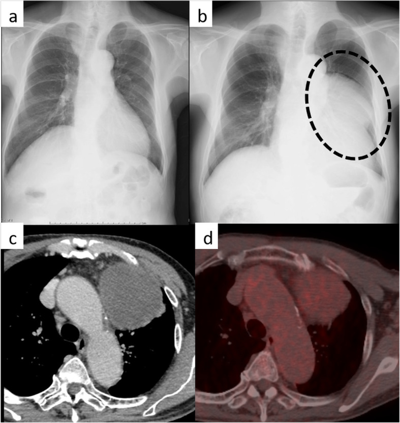
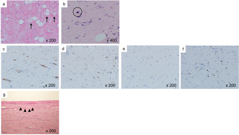
A 77-year-old man with a giant mass in the left thoracic cavity was referred to our hospital. This mass measured about 10 cm and occupied the left-sided mediastinum on a chest radiography; however, there was no abnormal finding on the previous year's chest radiography. Chest-enhanced computed tomography revealed a well-circumscribed 11-cm mass in the left-sided anterior mediastinum. Positron emission tomography showed accumulation of fluorodeoxyglucose uptake in this tumor (maximum standard uptake value = 3.3). The radiological findings of computed tomography and positron emission tomography indicated that this tumor was a benign or low-grade malignancy; therefore, the chest radiographic findings were difficult to explain. To explain this discrepancy and establish the diagnosis, tumor resection was performed via left posterolateral thoracotomy. Intraoperatively, the left phrenic nerve and pericardium were adhered tightly to the tumor, so we resected them. The tumor was well-circumscribed and fibrous; therefore, the initial diagnosis was solitary fibrous tumor. However, based on its histopathological and immunohistochemical patterns, the tumor was diagnosed as a sclerosing variant of well-differentiated liposarcoma. Five years postoperatively, the patient remains alive with no evidence of disease recurrence.
A well-differentiated liposarcoma is usually considered a low-grade malignancy. Nevertheless, the giant tumor in the present case appeared within 1 year. Thus, this was an extremely rare case of a sclerosing variant of well-differentiated liposarcoma with rapid growth.
纵隔脂肪肉瘤罕见,占纵隔肿瘤的比例不到1%。此外,快速生长的高分化脂肪肉瘤极为罕见。高分化脂肪肉瘤通常被认为是低级别恶性肿瘤。然而,我们报告了一例极为罕见的高分化脂肪肉瘤硬化型病例,该肿瘤在一年内迅速生长。
一名77岁男性因左胸腔巨大肿块被转诊至我院。该肿块大小约10厘米,胸部X线片显示占据左侧纵隔;然而,上一年的胸部X线片未发现异常。胸部增强计算机断层扫描显示左侧前纵隔有一个边界清晰的11厘米肿块。正电子发射断层扫描显示该肿瘤有氟脱氧葡萄糖摄取积聚(最大标准摄取值=3.3)。计算机断层扫描和正电子发射断层扫描的影像学表现表明该肿瘤为良性或低级别恶性肿瘤;因此,胸部X线片的表现难以解释。为了解释这种差异并明确诊断,通过左后外侧开胸术进行了肿瘤切除。术中,左膈神经和心包与肿瘤紧密粘连,因此我们将其切除。肿瘤边界清晰且质地坚韧;因此,初步诊断为孤立性纤维瘤。然而,根据其组织病理学和免疫组化模式,该肿瘤被诊断为高分化脂肪肉瘤硬化型。术后五年,患者存活,无疾病复发迹象。
高分化脂肪肉瘤通常被认为是低级别恶性肿瘤。然而,本例中的巨大肿瘤在1年内出现。因此,这是一例极为罕见的高分化脂肪肉瘤硬化型快速生长病例。