Department of Biomedical Physiology and Kinesiology, Simon Fraser University, 8888 University Dr, Burnaby, BC, V5A 1S6, Canada.
Behavioral & Cognitive Neuroscience Institute, Simon Fraser University, Burnaby, Canada.
Sci Rep. 2020 Jul 6;10(1):11067. doi: 10.1038/s41598-020-67507-3.
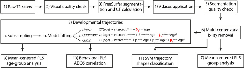
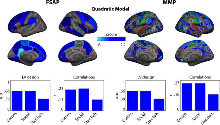
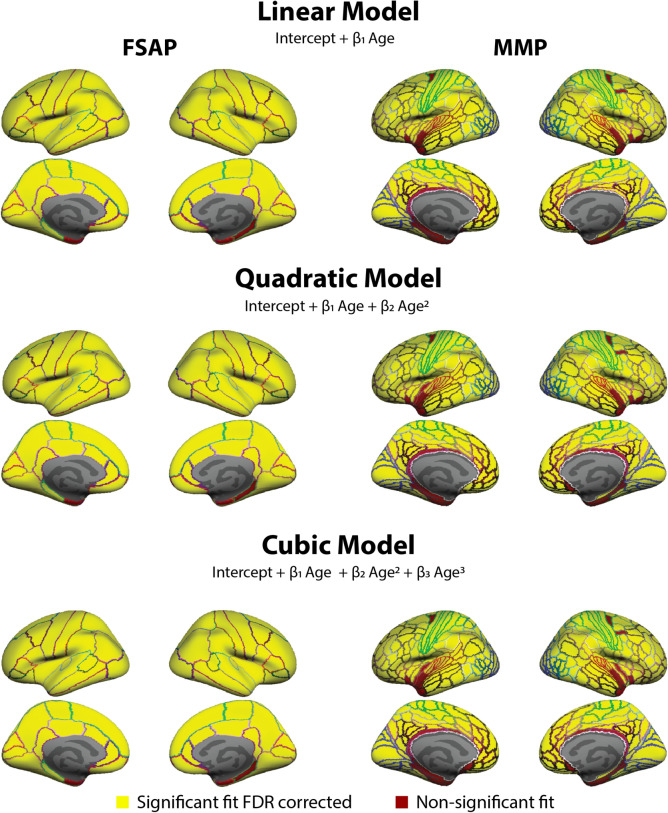
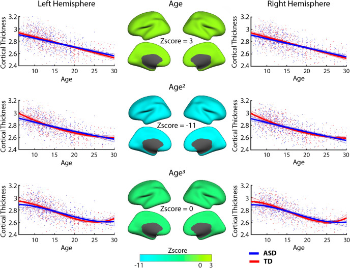
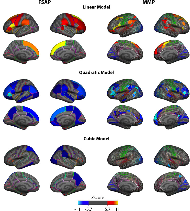
Recent longitudinal neuroimaging and neurophysiological studies have shown that tracking relative age-related changes in neural signals, rather than a static snapshot of a neural measure, could offer higher sensitivity for discriminating typically developing (TD) individuals from those with autism spectrum disorder (ASD). It is not clear, however, which aspects of age-related changes (trajectories) would be optimal for identifying atypical brain development in ASD. Using a large cross-sectional data set (Autism Brain Imaging Data Exchange [ABIDE] repository; releases I and II), we aimed to explore age-related changes in cortical thickness (CT) in TD and ASD populations (age range 6-30 years old). Cortical thickness was estimated from T1-weighted MRI images at three scales of spatial coarseness (three parcellations with different numbers of regions of interest). For each parcellation, three polynomial models of age-related changes in CT were tested. Specifically, to characterize alterations in CT trajectories, we compared the linear slope, curvature, and aberrancy of CT trajectories across experimental groups, which was estimated using linear, quadratic, and cubic polynomial models, respectively. Also, we explored associations between age-related changes with ASD symptomatology quantified as the Autism Diagnostic Observation Schedule (ADOS) scores. While no overall group differences in cortical thickness were observed across the entire age range, ASD and TD populations were different in terms of age-related changes, which were located primarily in frontal and tempo-parietal areas. These atypical age-related changes were also associated with ADOS scores in the ASD group and used to predict ASD from TD development. These results indicate that the curvature is the most reliable feature for localizing brain areas developmentally atypical in ASD with a more pronounced effect with symptomatology and is the most sensitive in predicting ASD development.
最近的纵向神经影像学和神经生理学研究表明,跟踪相对年龄相关的神经信号变化,而不是对神经测量值进行静态快照,可能会提高区分自闭症谱系障碍(ASD)患者和典型发育(TD)个体的敏感性。然而,尚不清楚年龄相关变化(轨迹)的哪些方面最适合识别 ASD 中的异常大脑发育。我们使用了一个大型的横断面数据集(自闭症脑成像数据交换[ABIDE]存储库;发布版 I 和 II),旨在探索 TD 和 ASD 人群中皮质厚度(CT)的年龄相关性变化(年龄范围为 6-30 岁)。从 T1 加权 MRI 图像中估算皮质厚度,在三个空间粗糙度尺度(具有不同感兴趣区域数量的三个分割)上进行。对于每个分割,测试了三种与年龄相关的 CT 变化的多项式模型。具体来说,为了描述 CT 轨迹的变化,我们比较了线性斜率、曲率和 CT 轨迹的异常,这些分别使用线性、二次和三次多项式模型进行估计。此外,我们还探索了与 ASD 症状相关的年龄相关变化之间的关联,该症状用自闭症诊断观察量表(ADOS)评分来量化。虽然在整个年龄范围内没有观察到皮质厚度的总体组间差异,但 ASD 和 TD 人群在年龄相关变化方面存在差异,这些差异主要位于额顶叶区域。这些非典型的年龄相关变化也与 ASD 组中的 ADOS 评分相关,并用于从 TD 发育中预测 ASD。这些结果表明,曲率是定位 ASD 中发育异常的脑区的最可靠特征,与症状的相关性更强,并且在预测 ASD 发育方面最敏感。