Andrews Derek S, Dakopolos Andrew J, Lee Joshua K, Heath Brianna, Cordero Devani, Solomon Marjorie, Amaral David G, Nordahl Christine Wu
Department of Psychiatry & Behavioral Sciences, the MIND Institute, University of California, Davis, California, USA.
A.A. Martinos Center for Biomedical Imaging, Massachusetts General Hospital, Boston, Massachusetts, USA.
Autism Res. 2025 Mar;18(3):486-497. doi: 10.1002/aur.3313. Epub 2025 Jan 30.
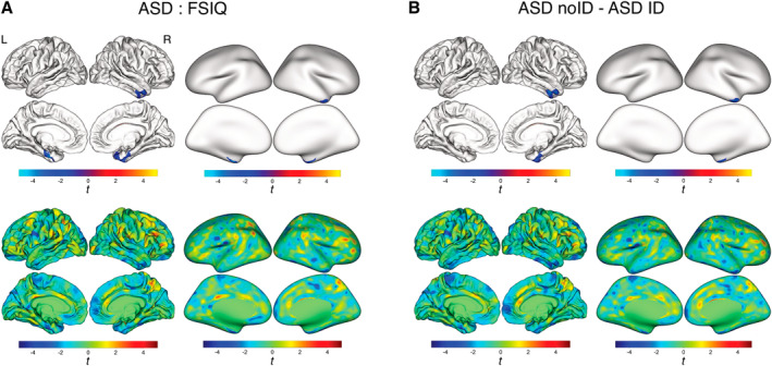
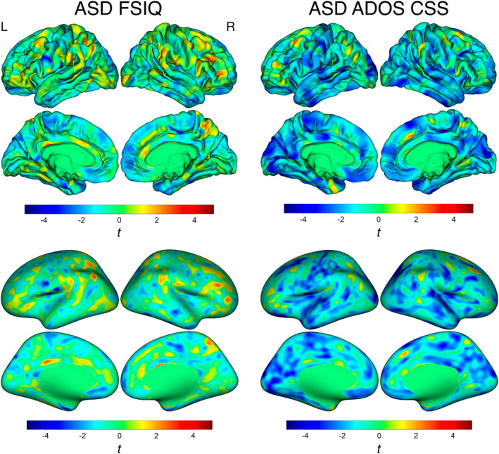
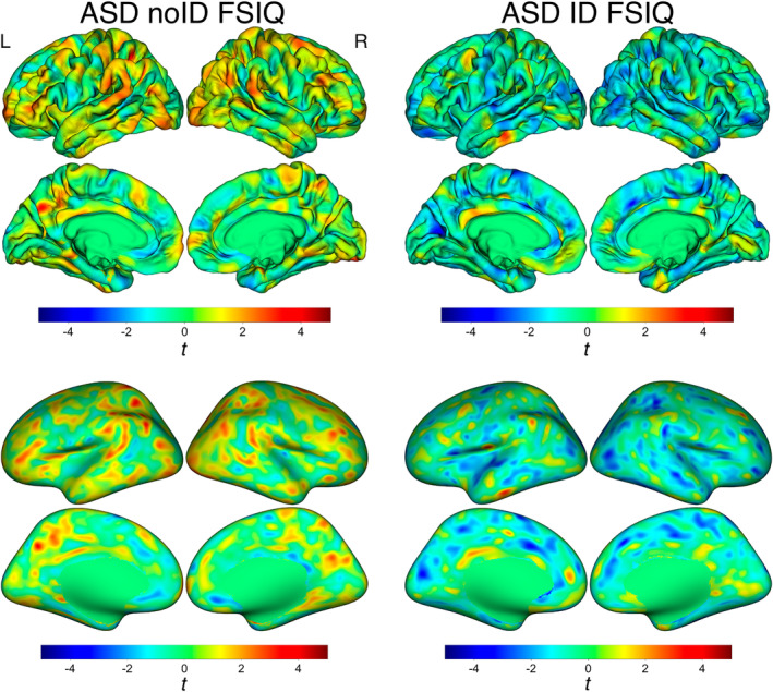
Of the 1 in 36 individuals in the United States who are diagnosed with autism spectrum disorder, nearly 40% also have intellectual disability (ID). The cortex has been widely implicated in neural processes underlying autistic behaviors as well as intellectual ability. Thus, neuroimaging features such as cortical thickness are of particular interest as a possible biomarkers of the condition. However, neuroimaging studies often fail to include autistic individuals with ID. As a result, there are few studies of cortical thickness in autistic individuals across the entire range of intellectual abilities. This study used MRI to evaluate cortical thickness in young autistic children (n = 88, mean age 5.37 years) with a large range of intellectual ability (IQ 19-133) as well as nonautistic, nondevelopmentally delayed (referred to here as typically developing [TD]) peers (n = 53, mean age 5.29 years). We first investigated associations between full scale IQ and cortical thickness in both autistic and TD children. Autistic children had significant negative associations (i.e., thinner cortex, higher IQ) in bilateral entorhinal cortex, right fusiform gyrus, superior, middle and inferior temporal gyri, and right temporal pole that were not present in TD children. Significantly thicker cortex was also observed in these regions for autistic children with ID (i.e., IQ ≤ 70) compared with those without. Last, given the reported correspondence between the severity of autism symptoms and intellectual ability, we compared cortical thickness associations with both IQ and ADOS Calibrated Severity Scores and found these patterns overlapped to a significant degree across the cortex.
在美国,每36个人中就有1人被诊断患有自闭症谱系障碍,其中近40%还伴有智力残疾(ID)。大脑皮层与自闭症行为以及智力所基于的神经过程密切相关。因此,诸如皮层厚度等神经影像学特征作为该病症可能的生物标志物备受关注。然而,神经影像学研究往往未纳入患有ID的自闭症个体。结果,针对不同智力水平的自闭症个体的皮层厚度研究很少。本研究使用磁共振成像(MRI)评估了智力水平差异较大(智商19 - 133)的自闭症幼儿(n = 88,平均年龄5.37岁)以及非自闭症、无发育迟缓(在此称为典型发育[TD])的同龄人(n = 53,平均年龄5.29岁)的皮层厚度。我们首先研究了自闭症儿童和TD儿童的全量表智商与皮层厚度之间的关联。自闭症儿童在双侧内嗅皮层、右侧梭状回、颞上回、颞中回、颞下回以及右侧颞极存在显著的负相关(即皮层越薄,智商越高),而TD儿童不存在这种情况。与无ID的自闭症儿童相比,患有ID(即智商≤70)的自闭症儿童在这些区域的皮层也明显更厚。最后,鉴于已报道的自闭症症状严重程度与智力之间的对应关系,我们比较了皮层厚度与智商以及自闭症诊断观察量表(ADOS)校准严重程度评分之间的关联,发现这些模式在整个皮层中在很大程度上重叠。