• 文献检索
  • 文档翻译
  • 深度研究
  • 学术资讯
  • Suppr Zotero 插件Zotero 插件
  • 邀请有礼
  • 套餐&价格
  • 历史记录
应用&插件
Suppr Zotero 插件Zotero 插件浏览器插件Mac 客户端Windows 客户端微信小程序
定价
高级版会员购买积分包购买API积分包
服务
文献检索文档翻译深度研究API 文档MCP 服务
关于我们
关于 Suppr公司介绍联系我们用户协议隐私条款
关注我们

Suppr 超能文献

核心技术专利:CN118964589B侵权必究
粤ICP备2023148730 号-1Suppr @ 2026

文献检索

告别复杂PubMed语法,用中文像聊天一样搜索,搜遍4000万医学文献。AI智能推荐,让科研检索更轻松。

立即免费搜索

文件翻译

保留排版,准确专业,支持PDF/Word/PPT等文件格式,支持 12+语言互译。

免费翻译文档

深度研究

AI帮你快速写综述,25分钟生成高质量综述,智能提取关键信息,辅助科研写作。

立即免费体验

应用弥散张量成像技术检测额颞叶痴呆亚型的皮质变化。

Using diffusion tensor imaging to detect cortical changes in fronto-temporal dementia subtypes.

机构信息

Nuffield Department of Clinical Neurosciences, University of Oxford, Oxford, UK.

Oxford Brain Diagnostics, Oxford Centre for Innovation, New Road, Oxford, OX1 1BY, UK.

出版信息

Sci Rep. 2020 Jul 8;10(1):11237. doi: 10.1038/s41598-020-68118-8.

DOI:10.1038/s41598-020-68118-8
PMID:32641807
原文链接:https://pmc.ncbi.nlm.nih.gov/articles/PMC7343779/
Abstract

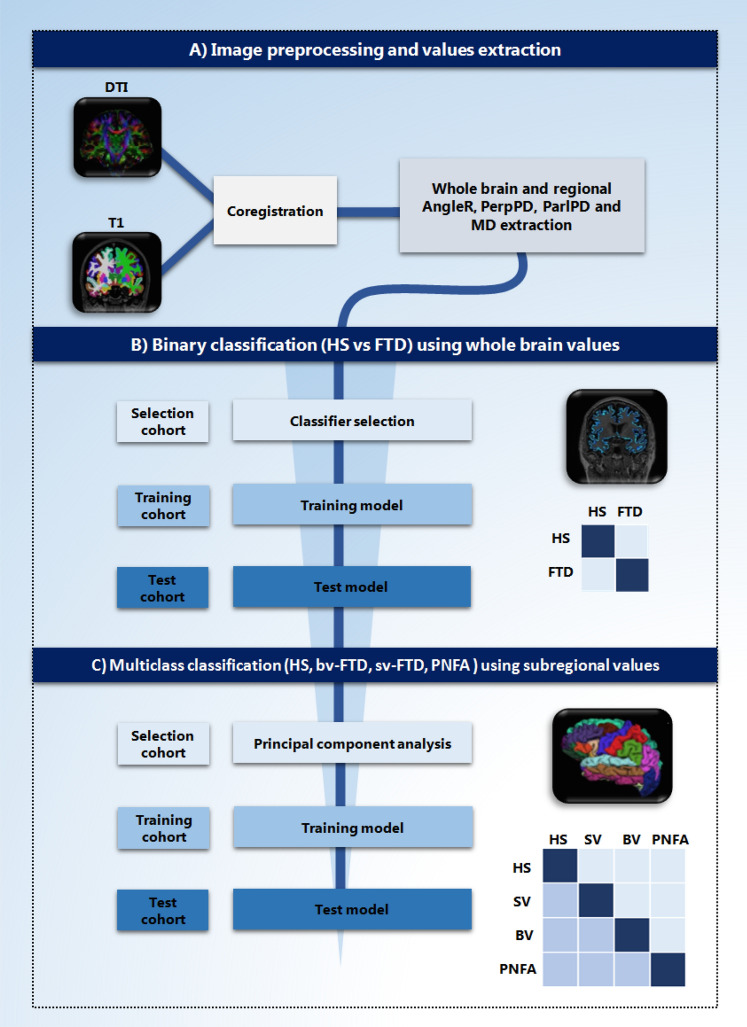
Fronto-temporal dementia (FTD) is a common type of presenile dementia, characterized by a heterogeneous clinical presentation that includes three main subtypes: behavioural-variant FTD, non-fluent/agrammatic variant primary progressive aphasia and semantic variant PPA. To better understand the FTD subtypes and develop more specific treatments, correct diagnosis is essential. This study aimed to test the discrimination power of a novel set of cortical Diffusion Tensor Imaging measures (DTI), on FTD subtypes. A total of 96 subjects with FTD and 84 healthy subjects (HS) were included in the study. A "selection cohort" was used to determine the set of features (measurements) and to use them to select the "best" machine learning classifier from a range of seven main models. The selected classifier was trained on a "training cohort" and tested on a third cohort ("test cohort"). The classifier was used to assess the classification power for binary (HS vs. FTD), and multiclass (HS and FTD subtypes) classification problems. In the binary classification, one of the new DTI features obtained the highest accuracy (85%) as a single feature, and when it was combined with other DTI features and two other common clinical measures (grey matter fraction and MMSE), obtained an accuracy of 88%. The new DTI features can distinguish between HS and FTD subgroups with an accuracy of 76%. These results suggest that DTI measures could support differential diagnosis in a clinical setting, potentially improve efficacy of new innovative drug treatments through effective patient selection, stratification and measurement of outcomes.

摘要

额颞叶痴呆(FTD)是一种常见的早发性痴呆症,其临床特征表现多样,包括三种主要亚型:行为变异型额颞叶痴呆、非流利/语法障碍型原发性进行性失语和语义变异型进行性失语。为了更好地理解 FTD 亚型并开发更具针对性的治疗方法,正确的诊断至关重要。本研究旨在测试一组新的皮质弥散张量成像(DTI)测量指标在 FTD 亚型中的区分能力。共有 96 名 FTD 患者和 84 名健康对照者(HS)纳入本研究。使用“选择队列”来确定特征(测量)集,并使用它们从七种主要模型中选择“最佳”机器学习分类器。选择的分类器在“训练队列”上进行训练,并在第三个队列(“测试队列”)上进行测试。该分类器用于评估二进制(HS 与 FTD)和多类(HS 和 FTD 亚型)分类问题的分类能力。在二进制分类中,一种新的 DTI 特征的准确性最高(85%),作为单一特征,当与其他 DTI 特征和另外两个常见的临床指标(灰质分数和 MMSE)结合使用时,准确性达到 88%。新的 DTI 特征可以区分 HS 和 FTD 亚组,准确率为 76%。这些结果表明,DTI 测量值可能支持临床环境中的鉴别诊断,通过有效选择患者、分层和测量结果,可能会提高新型创新药物治疗的疗效。

https://cdn.ncbi.nlm.nih.gov/pmc/blobs/478d/7343779/8b1e21b6170b/41598_2020_68118_Fig2_HTML.jpg
https://cdn.ncbi.nlm.nih.gov/pmc/blobs/478d/7343779/59692b511846/41598_2020_68118_Fig1_HTML.jpg
https://cdn.ncbi.nlm.nih.gov/pmc/blobs/478d/7343779/8b1e21b6170b/41598_2020_68118_Fig2_HTML.jpg
https://cdn.ncbi.nlm.nih.gov/pmc/blobs/478d/7343779/59692b511846/41598_2020_68118_Fig1_HTML.jpg
https://cdn.ncbi.nlm.nih.gov/pmc/blobs/478d/7343779/8b1e21b6170b/41598_2020_68118_Fig2_HTML.jpg

相似文献

1
Using diffusion tensor imaging to detect cortical changes in fronto-temporal dementia subtypes.应用弥散张量成像技术检测额颞叶痴呆亚型的皮质变化。
Sci Rep. 2020 Jul 8;10(1):11237. doi: 10.1038/s41598-020-68118-8.
2
Intracortical diffusion tensor imaging signature of microstructural changes in frontotemporal lobar degeneration.皮质内弥散张量成像对额颞叶变性的结构变化特征。
Alzheimers Res Ther. 2021 Oct 22;13(1):180. doi: 10.1186/s13195-021-00914-4.
3
Machine learning based hierarchical classification of frontotemporal dementia and Alzheimer's disease.基于机器学习的额颞叶痴呆和阿尔茨海默病的分层分类。
Neuroimage Clin. 2019;23:101811. doi: 10.1016/j.nicl.2019.101811. Epub 2019 Apr 3.
4
The power of neuroimaging biomarkers for screening frontotemporal dementia.神经影像学生物标志物在筛查额颞叶痴呆方面的作用。
Hum Brain Mapp. 2014 Sep;35(9):4827-40. doi: 10.1002/hbm.22515. Epub 2014 Mar 31.
5
Altered speech-related cortical network in frontotemporal dementia.额颞叶痴呆患者言语相关皮质网络的改变。
Brain Stimul. 2020 May-Jun;13(3):765-773. doi: 10.1016/j.brs.2020.02.029. Epub 2020 Feb 26.
6
Multiparametric computer-aided differential diagnosis of Alzheimer's disease and frontotemporal dementia using structural and advanced MRI.使用结构和高级磁共振成像对阿尔茨海默病和额颞叶痴呆进行多参数计算机辅助鉴别诊断。
Eur Radiol. 2017 Aug;27(8):3372-3382. doi: 10.1007/s00330-016-4691-x. Epub 2016 Dec 16.
7
Connectivity-based characterisation of subcortical grey matter pathology in frontotemporal dementia and ALS: a multimodal neuroimaging study.基于连接性的额颞叶痴呆和肌萎缩侧索硬化症皮质下灰质病理特征:一项多模态神经影像学研究。
Brain Imaging Behav. 2018 Dec;12(6):1696-1707. doi: 10.1007/s11682-018-9837-9.
8
Neuroimaging in frontotemporal dementia.额颞叶痴呆的神经影像学。
Int Rev Psychiatry. 2013 Apr;25(2):221-9. doi: 10.3109/09540261.2013.778822.
9
Defining Neuropsychiatric Inventory scale differences across frontotemporal dementia syndromes.明确额颞叶痴呆综合征之间的神经精神科问卷量表差异。
Psychogeriatrics. 2019 Jan;19(1):32-37. doi: 10.1111/psyg.12358. Epub 2018 Aug 2.
10
Differentiation between Subtypes of Primary Progressive Aphasia by Using Cortical Thickness and Diffusion-Tensor MR Imaging Measures.利用皮质厚度和弥散张量磁共振成像测量指标对原发性进行性失语症亚型进行鉴别。
Radiology. 2015 Jul;276(1):219-27. doi: 10.1148/radiol.15141869. Epub 2015 Feb 26.

引用本文的文献

1
Predicting progression from MCI to dementia using cortical disarray measurement from diffusion MRI.利用扩散磁共振成像测量皮质紊乱来预测从轻度认知障碍进展为痴呆症。
Alzheimers Dement. 2025 May;21(5):e70310. doi: 10.1002/alz.70310.
2
Cortical Diffusivity, a Biomarker for Early Neuronal Damage, Is Associated with Amyloid-β Deposition: A Pilot Study.皮质扩散率,一种早期神经元损伤的生物标志物,与β淀粉样蛋白沉积相关:一项初步研究。
Cells. 2025 Jan 21;14(3):155. doi: 10.3390/cells14030155.
3
Advanced Magnetic Resonance Imaging for Early Diagnosis and Monitoring of Movement Disorders.

本文引用的文献

1
Neural Correlates for Apathy: Frontal-Prefrontal and Parietal Cortical- Subcortical Circuits.冷漠的神经关联:额叶-前额叶和顶叶皮质-皮质下回路
Front Aging Neurosci. 2016 Dec 9;8:289. doi: 10.3389/fnagi.2016.00289. eCollection 2016.
用于运动障碍早期诊断和监测的先进磁共振成像
Brain Sci. 2025 Jan 16;15(1):79. doi: 10.3390/brainsci15010079.
4
Genetic Associations Among Inflammation, White Matter Architecture, and Extracellular Free Water.炎症、白质结构与细胞外游离水之间的遗传关联
Hum Brain Mapp. 2025 Jan;46(1):e70101. doi: 10.1002/hbm.70101.
5
Clinical utility of diffusion MRI-derived measures of cortical microstructure in a real-world memory clinic setting.弥散磁共振成像衍生的皮质微结构测量在真实世界记忆门诊中的临床应用。
Ann Clin Transl Neurol. 2024 Aug;11(8):1964-1976. doi: 10.1002/acn3.52097. Epub 2024 Jul 24.
6
Differential diagnosis of frontotemporal dementia subtypes with explainable deep learning on structural MRI.基于结构磁共振成像的可解释深度学习对额颞叶痴呆亚型的鉴别诊断
Front Neurosci. 2024 Feb 7;18:1331677. doi: 10.3389/fnins.2024.1331677. eCollection 2024.
7
In vivo cortical diffusion imaging relates to Alzheimer's disease neuropathology.体内皮质扩散成像与阿尔茨海默病神经病理学有关。
Alzheimers Res Ther. 2023 Oct 4;15(1):165. doi: 10.1186/s13195-023-01309-3.
8
Multiclass characterization of frontotemporal dementia variants via multimodal brain network computational inference.通过多模态脑网络计算推理对额颞叶痴呆变体进行多类别特征描述。
Netw Neurosci. 2023 Jan 1;7(1):322-350. doi: 10.1162/netn_a_00285. eCollection 2023.
9
White matter microstructure alterations in frontotemporal dementia: Phenotype-associated signatures and single-subject interpretation.额颞叶痴呆的脑白质微观结构改变:表型相关特征和单个体解释。
Brain Behav. 2022 Feb;12(2):e2500. doi: 10.1002/brb3.2500. Epub 2022 Jan 24.
10
Intracortical diffusion tensor imaging signature of microstructural changes in frontotemporal lobar degeneration.皮质内弥散张量成像对额颞叶变性的结构变化特征。
Alzheimers Res Ther. 2021 Oct 22;13(1):180. doi: 10.1186/s13195-021-00914-4.