Huizer Karin, Sacchetti Andrea, Swagemakers Sigrid, van der Spek Peter J, Dik Wim, Mustafa Dana A, Kros Johan M
Laboratory for Tumor Immuno-Pathology, Erasmus Medical Center, Rotterdam, The Netherlands.
Department of Pathology and Clinical Bio-Informatics, Erasmus Medical Center, Rotterdam, The Netherlands.
Neurooncol Adv. 2020 Apr 1;2(1):vdaa040. doi: 10.1093/noajnl/vdaa040. eCollection 2020 Jan-Dec.
In order to identify suitable therapeutic targets for glioma anti-angiogenic therapy, the process of neovascularization mediated by circulating angiogenic cells (CACs) needs to be scrutinized.
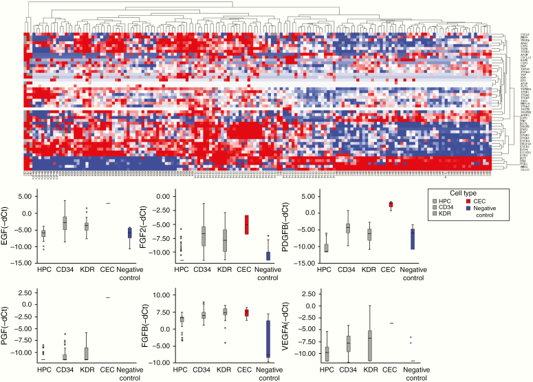
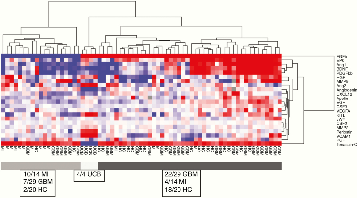
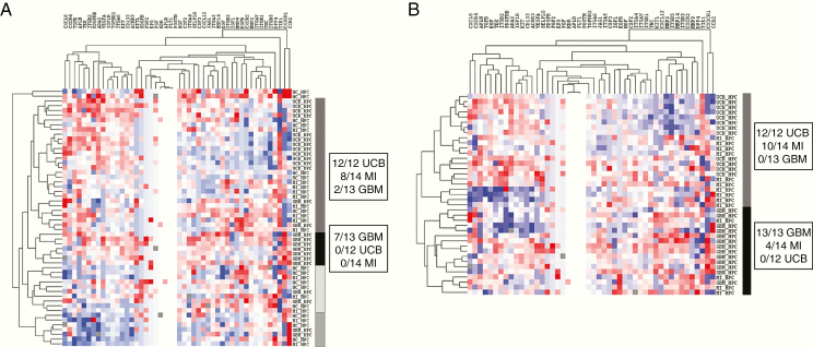
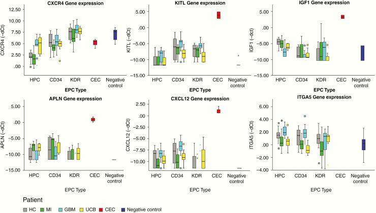
In the present study, we compared the expression of neovascularization-related genes by 3 circulating CAC subsets (hematopoietic progenitor cells [HPCs], CD34, and KDR cells; internal controls: peripheral blood mononuclear cells and circulating endothelial cells) of treatment-naïve patients with glioblastoma (GBM) to those of patients undergoing reactive neovascularization (myocardial infarction (MI). CACs from umbilical cord (representing developmental neovascularization) and healthy subjects served as controls. Fluorescent-activated cell sorting was used to isolate CACs, RT-PCR to determine the expression levels of a panel of 48 neovascularization-related genes, and Luminex assays to measure plasma levels of 21 CAC-related circulating molecules.
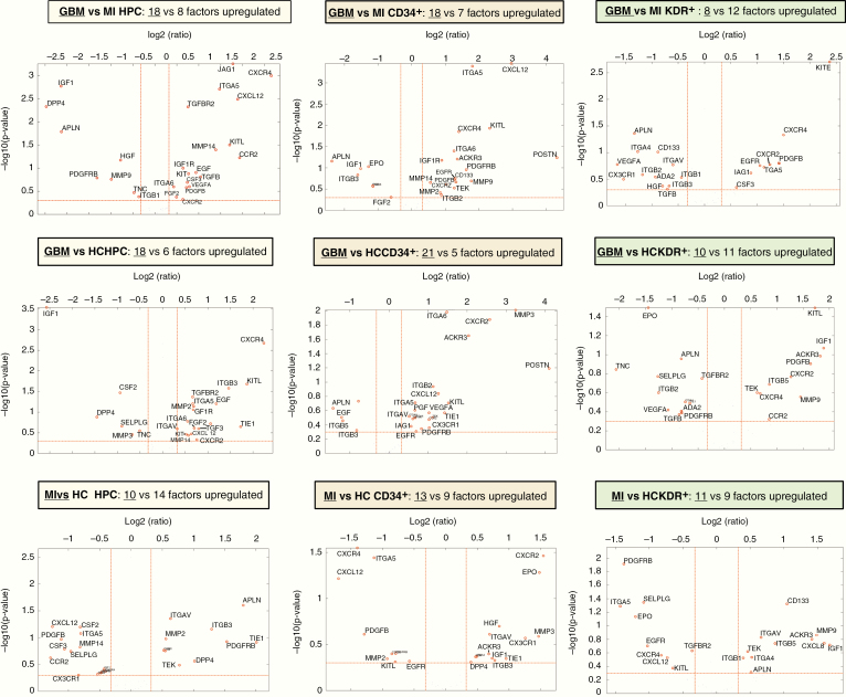
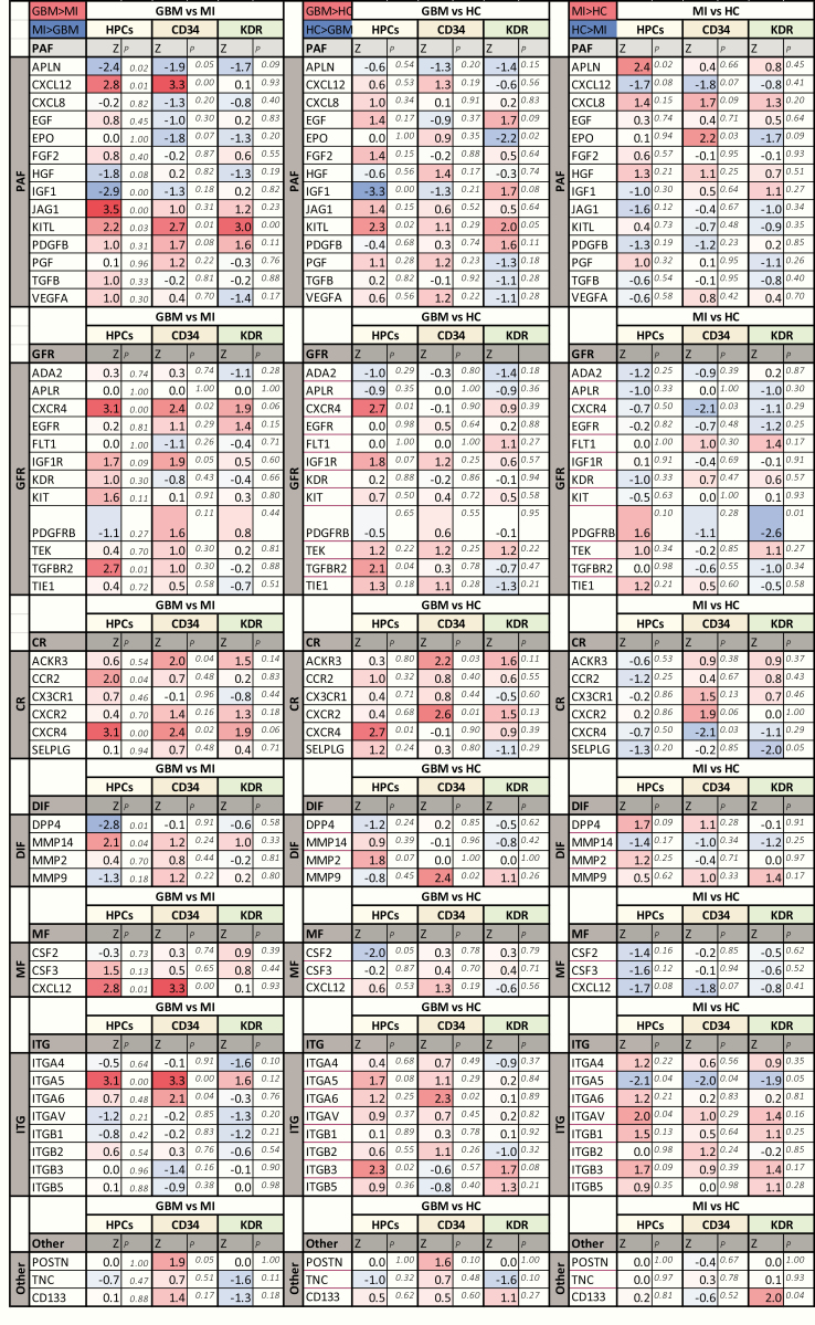
We found essential differences in gene expression between GBM and MI CACs. GBM CACs had a higher expression of proangiogenic factors (especially, , , and ), growth factor and chemotactic receptors (, , , and ), adhesion receptor monomers ( and ), and matricellular factor . In addition, we found major differences in the levels of neovascularization-related plasma factors. A strong positive correlation between plasma MMP9 levels and expression of in the CAC subset of HPCs was found in GBM patients.
Our findings indicate that CAC-mediated neovascularization in GBM is characterized by more efficient CAC homing to target tissue and a more potent proangiogenic response than in physiologic tissue repair in MI. Our findings can aid in selecting targets for therapeutic strategies acting against GBM-specific CACs.
为了确定胶质瘤抗血管生成治疗的合适靶点,需要仔细研究循环血管生成细胞(CACs)介导的新生血管形成过程。
在本研究中,我们比较了未经治疗的胶质母细胞瘤(GBM)患者的3种循环CAC亚群(造血祖细胞[HPCs]、CD34和KDR细胞;内部对照:外周血单核细胞和循环内皮细胞)与经历反应性新生血管形成的患者(心肌梗死[MI])的新生血管形成相关基因的表达。来自脐带(代表发育性新生血管形成)和健康受试者的CACs作为对照。使用荧光激活细胞分选技术分离CACs,采用逆转录聚合酶链反应(RT-PCR)测定一组48个新生血管形成相关基因的表达水平,并采用Luminex检测法测量21种与CAC相关的循环分子的血浆水平。
我们发现GBM和MI的CACs在基因表达上存在本质差异。GBM的CACs中促血管生成因子(特别是、、和)、生长因子和趋化因子受体(、、、和)、黏附受体单体(和)以及基质细胞因子的表达较高。此外,我们发现新生血管形成相关血浆因子水平存在重大差异。在GBM患者中,发现血浆基质金属蛋白酶9(MMP9)水平与HPCs的CAC亚群中的表达呈强正相关。
我们的研究结果表明,与MI的生理性组织修复相比,GBM中CAC介导的新生血管形成的特点是CAC更有效地归巢到靶组织,并且促血管生成反应更强。我们的研究结果有助于选择针对GBM特异性CACs的治疗策略的靶点。