Division of Clinical Geriatrics, Department of Neurobiology, Care Sciences and Society, Karolinska Institutet, Stockholm, Sweden.
Department of Biomedical Engineering and Health Systems (MTH), KTH Royal Institute of Technology, Stockholm, Sweden.
Aging (Albany NY). 2020 Jul 9;12(13):12622-12647. doi: 10.18632/aging.103623.
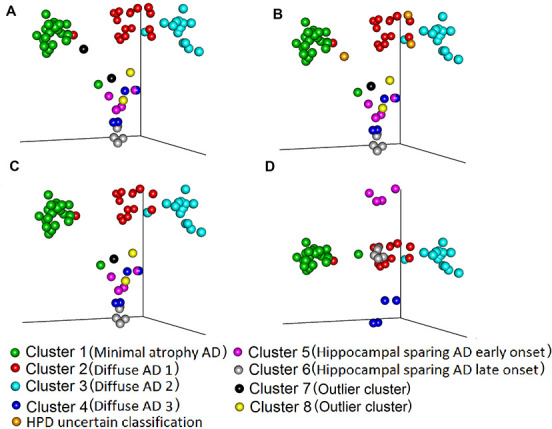
Tau pathology and brain atrophy are the closest correlate of cognitive decline in Alzheimer's disease (AD). Understanding heterogeneity and longitudinal progression of atrophy during the disease course will play a key role in understanding AD pathogenesis. We propose a framework for longitudinal clustering that simultaneously: 1) incorporates whole brain data, 2) leverages unequal visits per individual, 3) compares clusters with a control group, 4) allows for study confounding effects, 5) provides cluster visualization, 6) measures clustering uncertainty. We used amyloid-β positive AD and negative healthy subjects, three longitudinal structural magnetic resonance imaging scans (cortical thickness and subcortical volume) over two years. We found three distinct longitudinal AD brain atrophy patterns: one typical diffuse pattern (n=34, 47.2%), and two atypical patterns: minimal atrophy (n=23 31.9%) and hippocampal sparing (n=9, 12.5%). We also identified outliers (n=3, 4.2%) and observations with uncertain classification (n=3, 4.2%). The clusters differed not only in regional distributions of atrophy at baseline, but also longitudinal atrophy progression, age at AD onset, and cognitive decline. A framework for the longitudinal assessment of variability in cohorts with several neuroimaging measures was successfully developed. We believe this framework may aid in disentangling distinct subtypes of AD from disease staging.
tau 病理学和脑萎缩是阿尔茨海默病 (AD) 认知能力下降的最密切相关因素。了解疾病过程中萎缩的异质性和纵向进展将在理解 AD 发病机制方面发挥关键作用。我们提出了一种纵向聚类框架,该框架可以同时:1)纳入全脑数据,2)利用个体不等的访问次数,3)将聚类与对照组进行比较,4)允许研究混杂效应,5)提供聚类可视化,6)测量聚类不确定性。我们使用了淀粉样蛋白-β阳性 AD 和阴性健康受试者,在两年内进行了三次纵向结构磁共振成像扫描(皮质厚度和皮质下体积)。我们发现了三种不同的 AD 脑萎缩纵向模式:一种典型的弥漫性模式(n=34,47.2%),两种非典型模式:最小萎缩(n=23,31.9%)和海马体保留(n=9,12.5%)。我们还确定了离群值(n=3,4.2%)和分类不确定的观察值(n=3,4.2%)。这些聚类不仅在基线时的萎缩区域分布上有所不同,而且在纵向萎缩进展、AD 发病年龄和认知能力下降方面也有所不同。成功开发了一种用于对具有多种神经影像学测量的队列进行纵向评估的变异的框架。我们相信,该框架可能有助于将 AD 的不同亚型与疾病分期区分开来。