Departments of Pulmonology.
Radiology and Nuclear Medicine, Radboud University Medical Center, Nijmegen, The Netherlands.
J Bronchology Interv Pulmonol. 2021 Jan 1;28(1):60-69. doi: 10.1097/LBR.0000000000000697.
Bronchoscopic diagnosis of small peripheral lung lesions suspected of lung cancer remains a challenge. A successful endobronchial diagnosis comprises navigation, confirmation, and tissue acquisition. In all steps, 3-dimensional information is essential. Cone-beam computed tomography (CBCT) imaging can provide computed tomography information and 3-dimensional augmented fluoroscopy imaging. We assessed whether CBCT imaging can improve navigation and diagnosis of peripheral lesions by 2 clinical workflows with a cross-over design: (1) a primary CBCT and radial endobronchial ultrasound mini probe imaging-based approach and (2) a primary electromagnetic navigation (EMN) and radial endobronchial ultrasound mini probe imaging-based approach.
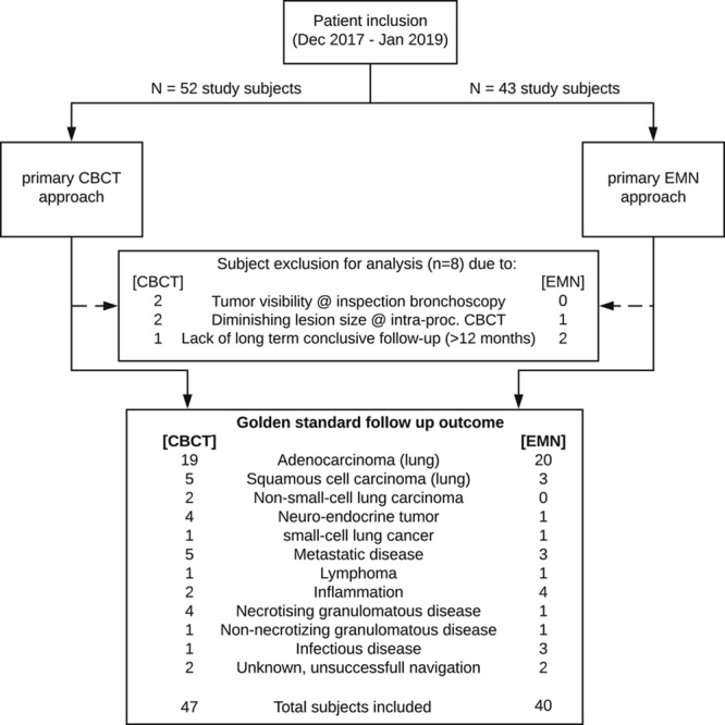
All patients with a peripheral lung lesion biopsy indication were eligible for study inclusion and randomly assigned to study arms. Commercially available equipment was used. The main study goals were to assess CBCT-confirmed navigation success and diagnostic accuracy. Surgery or unambiguous clinical follow-up served as the gold standard.
Eighty-seven patients with 107 lesions were included. Lesion mean longest axis size in the CBCT arm was 16.6 mm (n=47) and 14.2 mm in the EMN arm (n=40). The primary CBCT approach and primary EMN approach had 76.3% and 52.2% navigation success, respectively. Addition of EMN to the CBCT approach increased navigation success to 89.9%. Addition of CBCT imaging to the EMN approach significantly increased navigation success to 87.5% per lesion. The overall diagnostic accuracy per patient was significantly lower than the navigation success, being 72.4%.
CBCT imaging is a valuable addition to navigation bronchoscopy. Although overall navigation success was high, the diagnostic accuracy remains to be improved. Future research should focus on improving the tissue acquisition methodology.
疑似肺癌的小周边肺部病变的支气管镜诊断仍然是一个挑战。成功的支气管内诊断包括导航、确认和组织获取。在所有步骤中,三维信息都是必不可少的。锥形束计算机断层扫描(CBCT)成像可以提供计算机断层扫描信息和三维增强透视成像。我们通过交叉设计评估了 CBCT 成像是否可以通过两种临床工作流程改善外周病变的导航和诊断:(1)基于原发性 CBCT 和径向支气管内超声迷你探头成像的方法,(2)基于原发性电磁导航(EMN)和径向支气管内超声迷你探头成像的方法。
所有有外周肺病变活检指征的患者都符合研究纳入标准,并随机分配到研究组。使用商业上可获得的设备。主要研究目标是评估 CBCT 确认的导航成功率和诊断准确性。手术或明确的临床随访作为金标准。
87 例患者共 107 个病变纳入研究。CBCT 组病变最长轴尺寸的平均值为 16.6mm(n=47),EMN 组为 14.2mm(n=40)。原发性 CBCT 方法和原发性 EMN 方法的导航成功率分别为 76.3%和 52.2%。将 EMN 添加到 CBCT 方法中可将导航成功率提高到 89.9%。将 CBCT 成像添加到 EMN 方法中可显著提高每例病变的导航成功率至 87.5%。每位患者的整体诊断准确性明显低于导航成功率,为 72.4%。
CBCT 成像对导航支气管镜检查是一种有价值的补充。尽管总体导航成功率较高,但诊断准确性仍有待提高。未来的研究应集中于改进组织获取方法。