Styrvoky Kim, Schwalk Audra, Pham David, Madsen Kristine, Chiu Hsienchang T, Abu-Hijleh Muhanned
Division of Pulmonary and Critical Care Medicine, University of Texas Southwestern Medical Center, Dallas, TX, USA.
Department of Internal Medicine, University of Texas Southwestern Medical Center, Dallas, TX, USA.
J Thorac Dis. 2023 Sep 28;15(9):4836-4848. doi: 10.21037/jtd-23-587. Epub 2023 Aug 30.
Shape sensing robotic-assisted bronchoscopy (ssRAB) combined with radial endobronchial ultrasound (r-EBUS) and cone beam computed tomography (CBCT) is a newer diagnostic modality for the evaluation of pulmonary lesions. There is limited data describing the radiation dose of CBCT combined with ssRAB. The purpose of this study was to describe the technical factors associated with the use of CBCT combined with ssRAB to biopsy pulmonary lesions.
We conducted a single center, prospective observational study of patients undergoing ssRAB combined with fixed CBCT for the pulmonary lesion biopsy. We report our patient demographics, and pulmonary lesion and procedure characteristics.
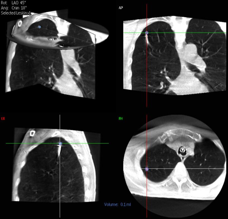
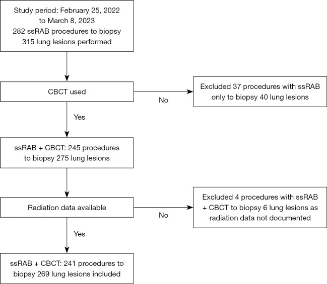
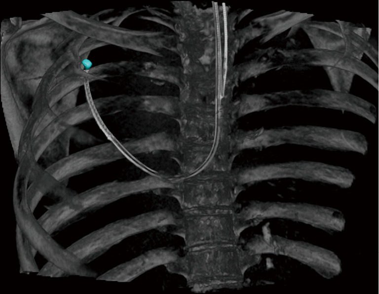
A total of 241 ssRAB procedures were performed to biopsy 269 pulmonary lesions. The mean lesion size was measured in the following dimensions: anteroposterior (18.0±8.8 mm), transverse (17.2±10.5 mm), and craniocaudal (17.7±10.2 mm). A mean of 1.5±0.7 (median: 1, range: 1-4) CBCT spins were performed. The mean total fluoroscopy time (FT) was 5.6±2.9 minutes. The mean radiation dose of cumulative air kerma (CAK) was 63.5±46.7 mGy and the mean cumulative dose area product (DAP) was 22.6±16.0 Gy·cm. Diagnostic yield calculated based on results at index bronchoscopy was 85.9%. There was a low rate of complications with 8 pneumothoraces (3.3%), 5 (2.1%) of which required chest tube placement.
We describe the use of ssRAB combined with CBCT to biopsy pulmonary lesions as a safe diagnostic modality with relatively low radiation dose that is potentially comparable to other image guided sampling modalities. Bronchoscopists should be cognizant of the radiation use during the procedure for both patient and staff safety.
形状感知机器人辅助支气管镜检查(ssRAB)联合径向支气管内超声(r-EBUS)和锥形束计算机断层扫描(CBCT)是评估肺部病变的一种较新的诊断方式。描述CBCT联合ssRAB辐射剂量的数据有限。本研究的目的是描述与使用CBCT联合ssRAB对肺部病变进行活检相关的技术因素。
我们对接受ssRAB联合固定CBCT进行肺部病变活检的患者进行了一项单中心前瞻性观察研究。我们报告了患者的人口统计学信息、肺部病变及操作特征。
共进行了241例ssRAB操作以对269个肺部病变进行活检。平均病变大小在以下维度测量:前后径(18.0±8.8毫米)、横径(17.2±10.5毫米)和头尾径(17.7±10.2毫米)。平均进行了1.5±0.7次(中位数:1,范围:1 - 4)CBCT旋转。平均总透视时间(FT)为5.6±2.9分钟。累积空气比释动能(CAK)的平均辐射剂量为63.5±46.7毫戈瑞,平均累积剂量面积乘积(DAP)为22.6±16.0戈瑞·厘米。根据初次支气管镜检查结果计算的诊断率为85.9%。并发症发生率较低,有8例气胸(3.3%),其中5例(2.1%)需要放置胸管。
我们描述了使用ssRAB联合CBCT对肺部病变进行活检是一种安全的诊断方式,辐射剂量相对较低,可能与其他影像引导采样方式相当。为了患者和工作人员的安全,支气管镜检查人员在操作过程中应注意辐射的使用。