From the Department of Radiology, Eye & ENT Hospital, Fudan University, Shanghai, China.
J Comput Assist Tomogr. 2021;45(1):135-141. doi: 10.1097/RCT.0000000000001065.
The purpose of this study was to explore the characteristic computed tomography (CT) and magnetic resonance (MR) features of small cell neuroendocrine carcinoma (SNEC) of paranasal sinuses.
Computed tomography (n = 8) and MR (n = 14) images and clinical findings from 14 patients with SNEC of paranasal sinuses were retrospectively reviewed.
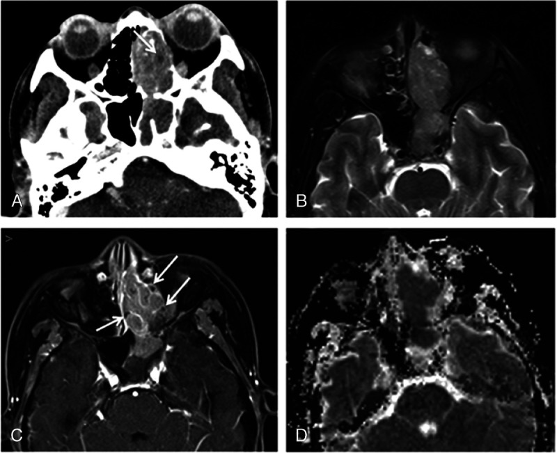
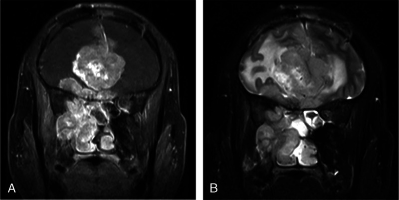
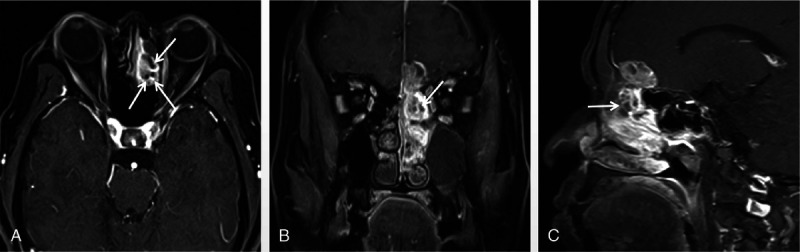
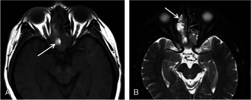
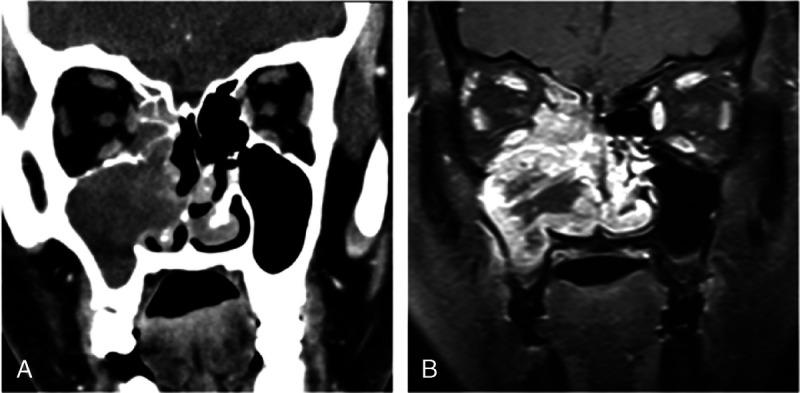
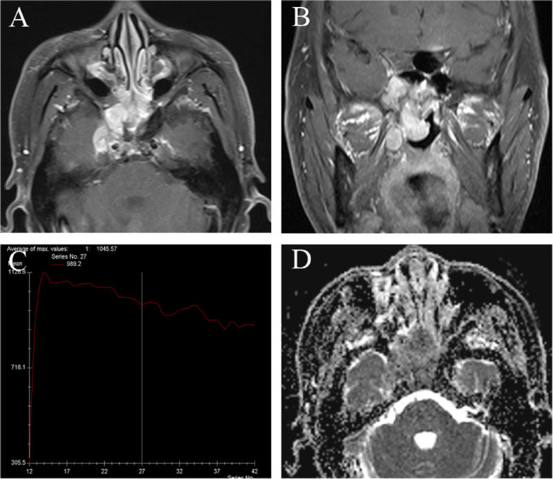
Eight lesions were located in the ethmoidal sinus, 4 in the maxillary sinus, and 2 in the sphenoid sinus. Small cell neuroendocrine carcinoma of the sphenoid sinus showed bilateral asymmetry patterns. On CT images, bony changes were visible in all 8 cases. On MR, 4 cases contained hemorrhage, and 10 cases contained cystic or necrotic areas. All cases demonstrated marked heterogeneous enhancement, with half showing a "cribriform-like" or "geographic" appearance. The nasal cavity was the most common site invaded by SNEC of paranasal sinuses, followed by the orbits. A time-signal intensity curve examination showed a washout-type pattern in all but 1 case. The mean ± SD apparent diffusion coefficient value was 0.702 ± 0.112 (×10-3 mm2/s). According to the Dulguerov staging system, 9 tumors were staged as N0 (1 T1, 1 T2, 5 T3, and 2 T4). The recurrence rate was 64.3%.
Some characteristics of radiological findings can provide important clues for preoperative diagnosis.
本研究旨在探讨发生于鼻窦的小细胞神经内分泌癌(SNEC)的 CT 和磁共振(MR)特征。
回顾性分析 14 例鼻窦 SNEC 患者的 CT(n=8)和 MR(n=14)图像及临床资料。
8 个病变位于筛窦,4 个位于上颌窦,2 个位于蝶窦。蝶窦 SNEC 呈双侧不对称性表现。CT 图像上,所有 8 例均可见骨改变。MR 上,4 例含出血,10 例含囊变或坏死区。所有病例均表现为明显不均匀强化,半数呈“筛孔样”或“地图样”外观。SNEC 最常侵犯鼻腔,其次为眼眶。除 1 例外,所有病例时间-信号强度曲线检查均呈流出型。表观弥散系数的平均值±标准差为 0.702±0.112(×10-3mm2/s)。根据 Dulguerov 分期系统,9 个肿瘤分期为 N0(1 个 T1、1 个 T2、5 个 T3 和 2 个 T4)。复发率为 64.3%。
一些影像学表现特征可为术前诊断提供重要线索。