Department of Radiology, Ningbo First Hospital, Ningbo, Zhejiang 315010, China.
Department of Imaging and Interventional Radiology, Faculty of Medicine, Prince of Wales Hospital, The Chinese University of Hong Kong, Hong Kong, China.
Chin Med J (Engl). 2020 Nov 20;133(22):2696-2702. doi: 10.1097/CM9.0000000000000919.
The importance of identifying osteoporotic vertebral endplate or/and cortex fracture (ECF), which primarily includes endplate fracture (EPF) and vertebral anterior cortex buckling, has been recognized. However, some old traumatic ECFs with healing process in the elderly may be mistaken as osteoporotic. This study analyzes the radiological features of traumatic EPF.
This was a retrospective analysis of 194 spine trauma patients with 263 vertebral fractures (mean age: 42.11 ± 9.82 years, 118 males and 76 females). All patients had traumatic EPF identified by X-ray/CT/MRI.
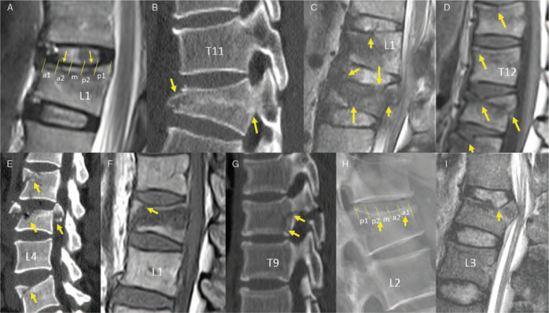
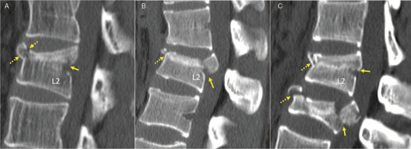
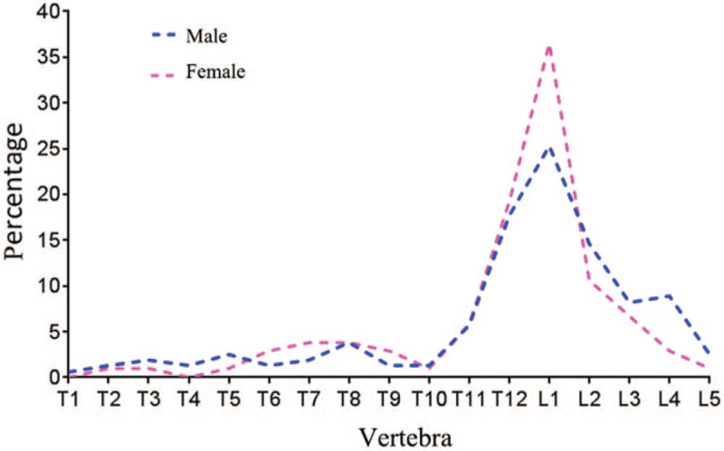
The involved vertebra was mostly L1 (29.7%), followed by T12 and L2. Except EPFs involved both superior and inferior endplates (12.6%), only 1.9% involved inferior endplate alone, with the majority involved superior endplate. If each endplate was divided into five segments of equal lengths (from anterior to posterior: a1, a2, m, p2, p1), the most depressed point of superior EPFs was mostly at segment-a2 (approximately 45%), followed by segment-a1 (approximately 20%) or segment-m (approximately 20%), and very rarely at segment-p1. The upper 1/3 of anterior vertebral wall was more likely to fracture, followed by middle 1/3 of anterior wall. For posterior vertebral wall fracture, 68.5% broke the bony wall surrounding the basivertebral vain. 58.6%, 30.0%, and 11.4% of vertebral fractures had <1/5, 1/5-1/3, and >1/3 vertebral body height loss. As the extent of vertebral height loss increased, the chance of having both superior and inferior EPFs also increased; however, the chance of having inferior EPF alone did not increase.
Traumatic EPF features are characterized, which may help the differentiation of traumatic and osteoporotic EPFs.
识别骨质疏松性椎体终板或/和皮质骨折(ECF)的重要性已得到认可,其中主要包括终板骨折(EPF)和椎体前皮质皱缩。然而,一些老年人中存在愈合过程的陈旧性创伤性 ECF 可能被误诊为骨质疏松性。本研究分析了外伤性 EPF 的影像学特征。
这是一项回顾性分析,共纳入 194 例脊柱创伤患者 263 个椎体骨折(平均年龄:42.11±9.82 岁,男性 118 例,女性 76 例)。所有患者均通过 X 线/CT/MRI 确诊为外伤性 EPF。
受累椎体以 L1 最常见(29.7%),其次是 T12 和 L2。除 EPF 累及上下终板(12.6%)外,仅 1.9%单独累及下终板,大部分累及上终板。如果将每个终板等分为 5 个长度相等的节段(从前向后:a1、a2、m、p2、p1),则上终板 EPF 的最凹陷点大多位于节段-a2(约 45%),其次是节段-a1(约 20%)或节段-m(约 20%),极少见位于节段-p1。前椎体壁的上 1/3 更容易骨折,其次是前壁的中 1/3。对于后椎体壁骨折,68.5%打破了围绕基底静脉的骨壁。58.6%、30.0%和 11.4%的椎体骨折椎体高度损失<1/5、1/5-1/3 和>1/3。随着椎体高度损失程度的增加,同时存在上下终板 EPF 的几率也增加;然而,单独存在下终板 EPF 的几率并未增加。
外伤性 EPF 的特征具有特征性,有助于外伤性和骨质疏松性 EPF 的鉴别。