• 文献检索
  • 文档翻译
  • 深度研究
  • 学术资讯
  • Suppr Zotero 插件Zotero 插件
  • 邀请有礼
  • 套餐&价格
  • 历史记录
应用&插件
Suppr Zotero 插件Zotero 插件浏览器插件Mac 客户端Windows 客户端微信小程序
定价
高级版会员购买积分包购买API积分包
服务
文献检索文档翻译深度研究API 文档MCP 服务
关于我们
关于 Suppr公司介绍联系我们用户协议隐私条款
关注我们

Suppr 超能文献

核心技术专利:CN118964589B侵权必究
粤ICP备2023148730 号-1Suppr @ 2026

文献检索

告别复杂PubMed语法,用中文像聊天一样搜索,搜遍4000万医学文献。AI智能推荐,让科研检索更轻松。

立即免费搜索

文件翻译

保留排版,准确专业,支持PDF/Word/PPT等文件格式,支持 12+语言互译。

免费翻译文档

深度研究

AI帮你快速写综述,25分钟生成高质量综述,智能提取关键信息,辅助科研写作。

立即免费体验

通过调整X射线束定位提高胸部正位X线片上骨质疏松性椎体畸形的检测率。

Improving osteoporotic vertebral deformity detection on chest frontal view radiograph by adjusted X-ray beam positioning.

作者信息

Du Er-Zhu, Liu Wei-Hong, Wáng Yì Xiáng J

机构信息

Department of Radiology, Dongguan Traditional Chinese Medicine Hospital, Dongguan, Guangdong province, China.

Department of Radiology, General Hospital of China Resources & Wuhan Iron and Steel Corporation, Wuhan, Hubei province, China.

出版信息

J Orthop Translat. 2021 May 5;28:169-178. doi: 10.1016/j.jot.2021.04.001. eCollection 2021 May.

DOI:10.1016/j.jot.2021.04.001
PMID:34036040
原文链接:https://pmc.ncbi.nlm.nih.gov/articles/PMC8121774/
Abstract

PURPOSE

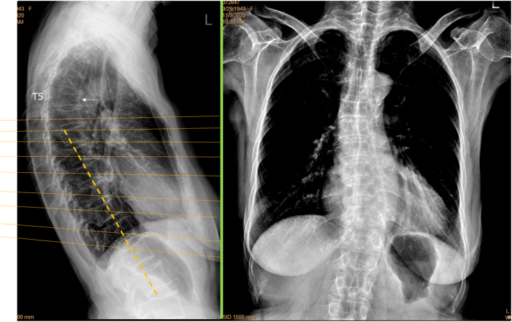
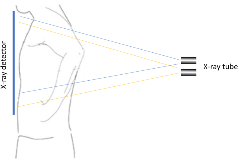
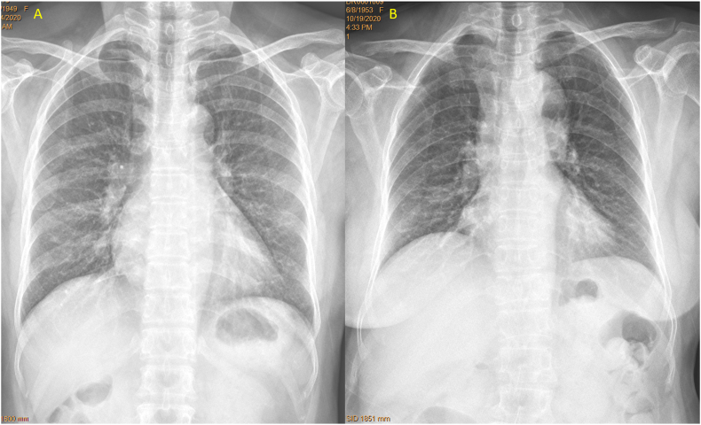
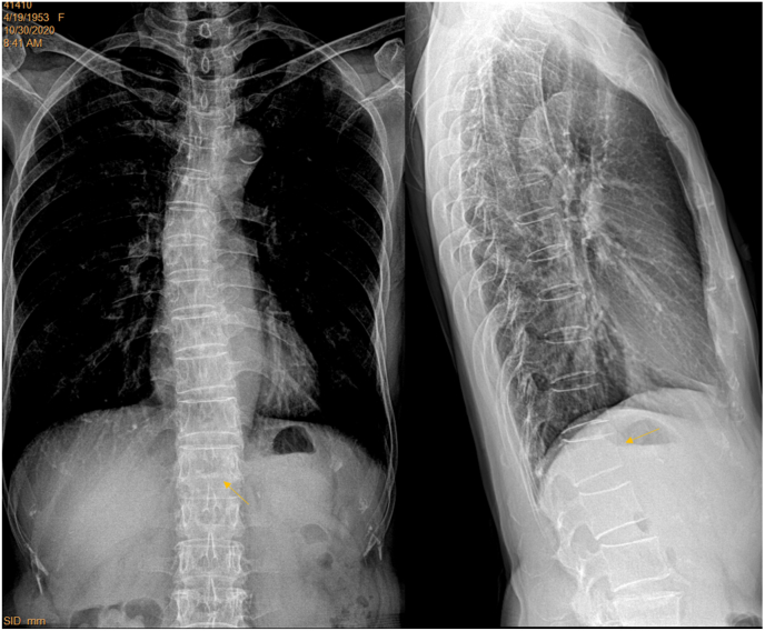
In clinics, chest frontal view radiograph (CFR) is often taken for patients suspected of respiratory diseases and for assessing the heart and big vessels. CFR can be utilised to opportunistically detect osteoporotic vertebral fracture (OVF). However, for standard CFR, the site of highest OVF prevalence, i.e., the thoracolumbar junction, is usually 'off-centre' to the X-ray beam focus. This study tested the hypothesis that, if a CRF is taken with approximately two vertebrae lower than the standard X-ray beam positioning, the visualization of thoraco-lumbar junction can be much improved.

MATERIALS

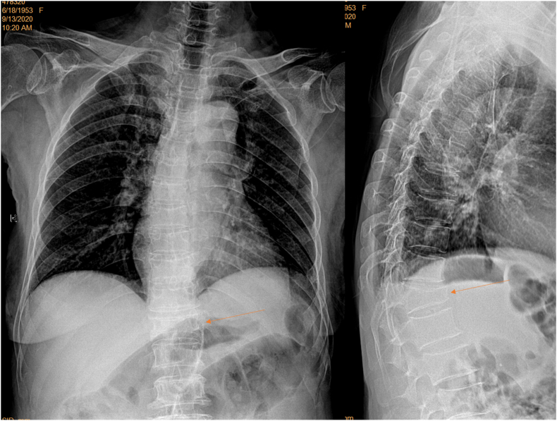
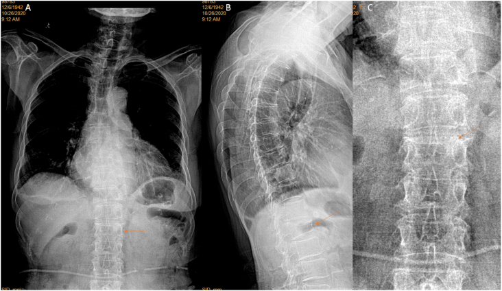
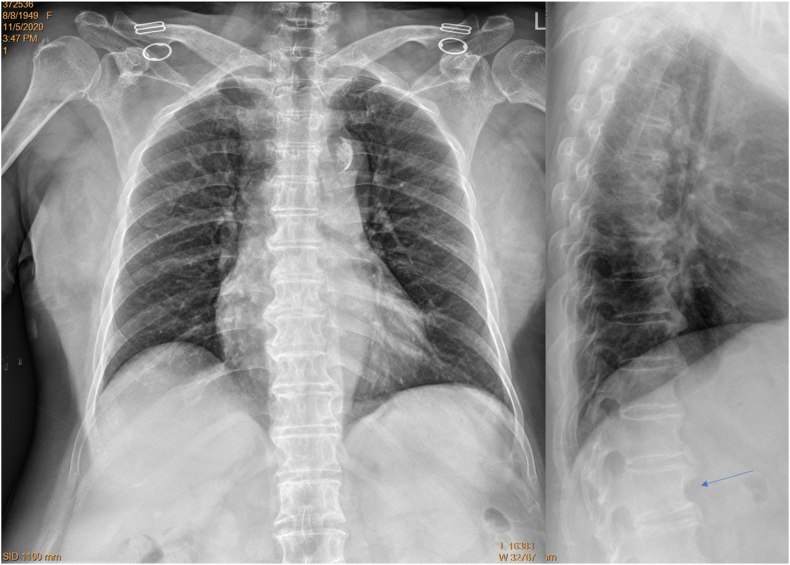
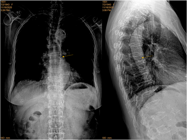
Four hospitals (A, B, C, D) provided 101 elderly women's digital CFRs with standard filming (28, 20, 24, and 21 cases respectively). Eighty four elderly female patients were prospectively recruited from hospitals-A and B, who were consecutive patients referred for chest radiograph with indications other than spine disorders. For theses prospective CFRs, the focus of X-ray beam was adjusted from towards vertebra T6 to towards T8, and standard lateral radiographs were obtained for reference. Visibility of spine and detectability of OVF were assessed on the CFRs. OVF was diagnosed based on chest lateral radiograph (CLR) after excluding other potential causes both radiographically and clinically.

RESULTS

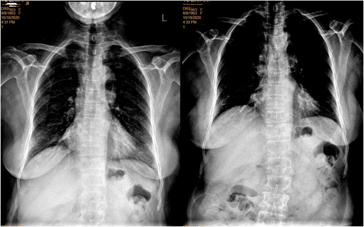
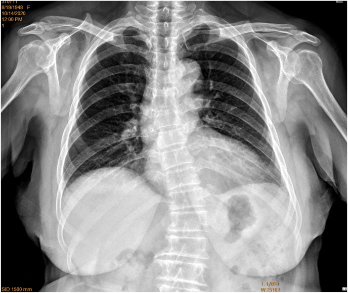
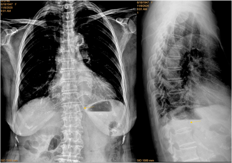
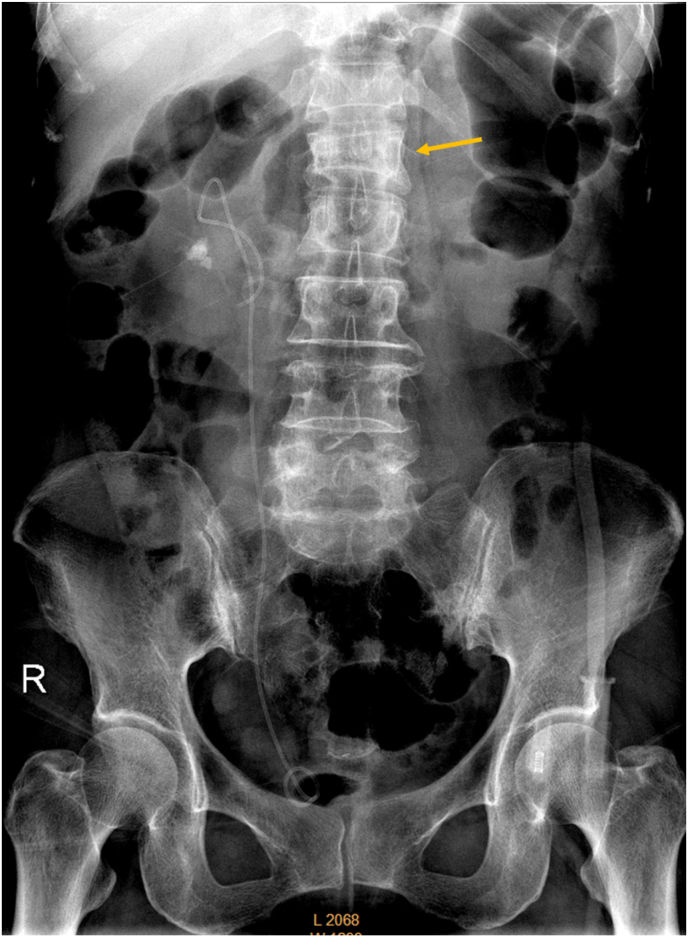
For standardly filmed CFR, spine readability was similar among those from Hospitals-A, B, and C, while performed less well for those from Hospital-D. With the prospective cases from Hospitals-A and B, spines readable to vertebra L1 level or lower increased from 48.2% for standard filming to 80.7% for adjusted filming. Spines with 'blurry' labelling decreased from 35.7% for standard filming to 15.7% for adjusted filming. For the 84 prospective cases, 42.9% (36/84) of the patients had OVF, and 26 cases of CLR positive cases were detected as having vertebral deformity on CFR. For minimal OVF cases (<20% height loss), 38% (5/13) were detected on CFR. Among 22 cases with apparent OVF (≥20% height loss), two cases were missed on CFR. False positivity was labelled in five cases, among them four cases had 'burry' spines.

CONCLUSION

CFR can help opportunistically detect OVF, which can be further improved if X-ray beam is adjusted to towards vertebra T8 instead of towards vertebra T6.

THE TRANSLATIONAL POTENTIAL OF THIS ARTICLE

This study confirms that CFR can help detect OVF opportunistically, and the visibility of the mid/lower thoracic spine and thoracolumbar junction can be much improved after minor adjustment of X-ray beam positioning. This study also suggests high positive rate of OVF in elderly Chinese female patients indicated for chest radiograph. Radiologists should be trained and sensitized in vertebral deformity identification on CFR as the clinical management can be improved by opportunistic detection of OVF.

https://cdn.ncbi.nlm.nih.gov/pmc/blobs/90a6/8121774/1c5cfef78dee/gr13.jpg
https://cdn.ncbi.nlm.nih.gov/pmc/blobs/90a6/8121774/ed82e6cff4cb/gr1.jpg
https://cdn.ncbi.nlm.nih.gov/pmc/blobs/90a6/8121774/0bc7f7fc37bb/gr2.jpg
https://cdn.ncbi.nlm.nih.gov/pmc/blobs/90a6/8121774/33f62f4ba022/gr3.jpg
https://cdn.ncbi.nlm.nih.gov/pmc/blobs/90a6/8121774/f18503109c59/gr4.jpg
https://cdn.ncbi.nlm.nih.gov/pmc/blobs/90a6/8121774/301990a3fe7f/gr5.jpg
https://cdn.ncbi.nlm.nih.gov/pmc/blobs/90a6/8121774/528a13bbabc5/gr6.jpg
https://cdn.ncbi.nlm.nih.gov/pmc/blobs/90a6/8121774/4c625c0f893f/gr7.jpg
https://cdn.ncbi.nlm.nih.gov/pmc/blobs/90a6/8121774/20c956789e9d/gr8.jpg
https://cdn.ncbi.nlm.nih.gov/pmc/blobs/90a6/8121774/08853edadbb9/gr9.jpg
https://cdn.ncbi.nlm.nih.gov/pmc/blobs/90a6/8121774/2d2d162f4daa/gr10.jpg
https://cdn.ncbi.nlm.nih.gov/pmc/blobs/90a6/8121774/e89981f03561/gr11.jpg
https://cdn.ncbi.nlm.nih.gov/pmc/blobs/90a6/8121774/c762df945ea3/gr12.jpg
https://cdn.ncbi.nlm.nih.gov/pmc/blobs/90a6/8121774/1c5cfef78dee/gr13.jpg
摘要

目的

在临床上,胸部正位X线片(CFR)常用于疑似患有呼吸系统疾病的患者以及用于评估心脏和大血管。CFR可用于机会性地检测骨质疏松性椎体骨折(OVF)。然而,对于标准的CFR,OVF患病率最高的部位,即胸腰段交界处,通常偏离X线束焦点。本研究检验了这样一个假设,即如果CFR拍摄时比标准X线束定位低大约两个椎体,胸腰段交界处的可视化效果会有很大改善。

材料

四家医院(A、B、C、D)提供了101例老年女性的数字化CFR,采用标准拍摄(分别为28例、20例、24例和21例)。从A医院和B医院前瞻性招募了84例老年女性患者,她们是因脊柱疾病以外的指征而被转诊进行胸部X线检查的连续患者。对于这些前瞻性CFR,将X线束焦点从T6椎体调整至T8椎体,并获得标准侧位X线片作为参考。在CFR上评估脊柱的可见性和OVF的可检测性。在排除其他影像学和临床潜在病因后,根据胸部侧位X线片(CLR)诊断OVF。

结果

对于标准拍摄的CFR,A、B和C医院的脊柱可读性相似,而D医院的表现较差。对于A医院和B医院的前瞻性病例,可读到L1椎体及以下的脊柱从标准拍摄时的48.2%增加到调整拍摄时的80.7%。标记“模糊”的脊柱从标准拍摄时的35.7%降至调整拍摄时的15.7%。对于84例前瞻性病例,42.9%(36/84)的患者患有OVF,26例CLR阳性病例在CFR上被检测出有椎体畸形。对于轻度OVF病例(椎体高度丢失<20%),38%(5/13)在CFR上被检测出。在22例明显OVF病例(椎体高度丢失≥20%)中,有2例在CFR上漏诊。有5例假阳性标记,其中4例脊柱“模糊”。

结论

CFR有助于机会性地检测OVF,如果将X线束调整至T8椎体而不是T6椎体,检测效果可进一步提高。

本文的转化潜力

本研究证实CFR有助于机会性地检测OVF,对X线束定位进行微小调整后,胸段中下部脊柱和胸腰段交界处的可视化效果可得到很大改善。本研究还提示,因胸部X线检查而就诊的中国老年女性患者中OVF的阳性率较高。放射科医生应接受培训并提高在CFR上识别椎体畸形的意识,因为机会性检测OVF可改善临床管理。

相似文献

1
Improving osteoporotic vertebral deformity detection on chest frontal view radiograph by adjusted X-ray beam positioning.通过调整X射线束定位提高胸部正位X线片上骨质疏松性椎体畸形的检测率。
J Orthop Translat. 2021 May 5;28:169-178. doi: 10.1016/j.jot.2021.04.001. eCollection 2021 May.
2
A software program for automated compressive vertebral fracture detection on elderly women's lateral chest radiograph: Ofeye 1.0.一款用于老年女性胸部侧位X光片自动检测椎体压缩性骨折的软件程序:Ofeye 1.0。
Quant Imaging Med Surg. 2022 Aug;12(8):4259-4271. doi: 10.21037/qims-22-433.
3
CT detects more osteoporotic endplate depressions than radiograph: a descriptive comparison of 76 vertebrae.CT 比 X 线更能检测到骨质疏松性终板凹陷:76 个椎体的描述性比较。
Osteoporos Int. 2022 Jul;33(7):1569-1577. doi: 10.1007/s00198-022-06391-1. Epub 2022 Apr 4.
4
Interpretation of osteoporotic vertebral deformity on frontal view radiographs of the chest and abdomen: a pictorial review.胸部和腹部正位X线片上骨质疏松性椎体畸形的解读:图文综述
Quant Imaging Med Surg. 2021 Jan;11(1):423-442. doi: 10.21037/qims-2020-28.
5
Recognizing osteoporotic vertebral deformity on frontal view radiograph: a cohort analysis and a pictorial review.基于队列分析和图像回顾的正位 X 线片上骨质疏松性椎体变形的识别。
Arch Osteoporos. 2020 Mar 6;15(1):41. doi: 10.1007/s11657-020-00716-5.
6
Symptomatic relevance of intravertebral cleft in patients with osteoporotic vertebral fracture.骨质疏松性椎体骨折患者椎体内裂隙的症状相关性。
J Neurosurg Spine. 2010 Aug;13(2):267-75. doi: 10.3171/2010.3.SPINE09364.
7
Radiological diagnosis of prevalent osteoporotic vertebral fracture on radiographs: an interim consensus from a group of experts of the ESSR osteoporosis and metabolism subcommittee.放射学诊断常见的骨质疏松性椎体骨折:ESSR 骨质疏松和代谢小组委员会专家组的临时共识。
Skeletal Radiol. 2024 Dec;53(12):2563-2574. doi: 10.1007/s00256-024-04678-4. Epub 2024 Apr 25.
8
The variability of vertebral body volume and pain associated with osteoporotic vertebral fractures: conservative treatment versus percutaneous transpedicular vertebroplasty.椎体体积变异性与骨质疏松性椎体骨折相关疼痛:保守治疗与经皮椎弓根椎体成形术对比
Int Orthop. 2017 May;41(5):963-968. doi: 10.1007/s00264-017-3409-2. Epub 2017 Feb 4.
9
Much lower prevalence and severity of radiographic osteoporotic vertebral fracture in elderly Hong Kong Chinese women than in age-matched Rome Caucasian women: a cross-sectional study.与年龄匹配的罗马白人女性相比,老年香港华裔女性的放射性骨质疏松性椎体骨折的患病率和严重程度要低得多:一项横断面研究。
Arch Osteoporos. 2021 Nov 16;16(1):174. doi: 10.1007/s11657-021-00987-6.
10
'Healthier Chinese spine': an update of osteoporotic fractures in men (MrOS) and in women (MsOS) Hong Kong spine radiograph studies.“更健康的中国脊柱”:香港脊柱X光片研究中男性骨质疏松性骨折(MrOS)和女性骨质疏松性骨折(MsOS)的最新情况
Quant Imaging Med Surg. 2022 Mar;12(3):2090-2105. doi: 10.21037/qims-2021-07.

引用本文的文献

1
Vertebral fracture severity assessment on anteroposterior radiographs with a new semi-quantitative technique.基于新半定量技术的前后位 X 射线片椎体骨折严重程度评估。
Osteoporos Int. 2024 May;35(5):831-839. doi: 10.1007/s00198-024-07024-5. Epub 2024 Jan 31.
2
An update of our understanding of radiographic diagnostics for prevalent osteoporotic vertebral fracture in elderly women.老年女性常见骨质疏松性椎体骨折影像学诊断的认识更新
Quant Imaging Med Surg. 2022 Jul;12(7):3495-3514. doi: 10.21037/qims-22-360.
3
'Healthier Chinese spine': an update of osteoporotic fractures in men (MrOS) and in women (MsOS) Hong Kong spine radiograph studies.

本文引用的文献

1
Interpretation of osteoporotic vertebral deformity on frontal view radiographs of the chest and abdomen: a pictorial review.胸部和腹部正位X线片上骨质疏松性椎体畸形的解读:图文综述
Quant Imaging Med Surg. 2021 Jan;11(1):423-442. doi: 10.21037/qims-2020-28.
2
The role of radiography in the study of spinal disorders.放射照相术在脊柱疾病研究中的作用。
Quant Imaging Med Surg. 2020 Dec;10(12):2322-2355. doi: 10.21037/qims-20-1014.
3
Elderly men have much lower vertebral fracture risk than elderly women even at advanced age: the MrOS and MsOS (Hong Kong) year 14 follow-up radiology results.
“更健康的中国脊柱”:香港脊柱X光片研究中男性骨质疏松性骨折(MrOS)和女性骨质疏松性骨折(MsOS)的最新情况
Quant Imaging Med Surg. 2022 Mar;12(3):2090-2105. doi: 10.21037/qims-2021-07.
即使在高龄时,老年男性的椎体骨折风险也远低于老年女性:MrOS 和 MsOS(香港)研究 14 年随访的影像学结果。
Arch Osteoporos. 2020 Nov 7;15(1):176. doi: 10.1007/s11657-020-00845-x.
4
Diagnosis and grading of radiographic osteoporotic vertebral deformity by general radiologists after a brief self-learning period.普通放射科医生在经过简短的自我学习期后对骨质疏松性椎体畸形进行影像学诊断和分级。
J Thorac Dis. 2020 Sep;12(9):4702-4710. doi: 10.21037/jtd-20-2379.
5
Chinese expert consensus on the diagnosis of osteoporosis by imaging and bone mineral density.中国骨质疏松症影像学与骨密度诊断专家共识
Quant Imaging Med Surg. 2020 Oct;10(10):2066-2077. doi: 10.21037/qims-2020-16.
6
Osteoporotic Vertebral Deformity: Radiological Appearances and Their Association With a History of Trauma and the Risk of Further Fragility Fracture.骨质疏松性椎体畸形:影像学表现及其与创伤史和进一步脆性骨折风险的关联。
Can Assoc Radiol J. 2021 Aug;72(3):585. doi: 10.1177/0846537120958471. Epub 2020 Sep 16.
7
Underreporting characteristics of osteoporotic vertebral fracture in back pain clinic patients of a tertiary hospital in China.中国一家三级医院背痛门诊患者骨质疏松性椎体骨折的漏报特征
J Orthop Translat. 2019 Nov 9;23:152-158. doi: 10.1016/j.jot.2019.10.007. eCollection 2020 Jul.
8
Vertebral Fractures: Which Radiological Criteria Are Better Associated With the Clinical Course of Osteoporosis?椎体骨折:哪些影像学标准与骨质疏松症的临床病程相关性更好?
Can Assoc Radiol J. 2021 Feb;72(1):150-158. doi: 10.1177/0846537120943529. Epub 2020 Aug 5.
9
The Belgian Bone Club 2020 guidelines for the management of osteoporosis in postmenopausal women.比利时骨俱乐部 2020 年绝经后妇女骨质疏松症管理指南。
Maturitas. 2020 Sep;139:69-89. doi: 10.1016/j.maturitas.2020.05.006. Epub 2020 May 16.
10
Radiological features of traumatic vertebral endplate fracture: an analysis of 194 cases with 263 vertebral fractures.创伤性椎体终板骨折的放射学特征:263 个椎体骨折的 194 例分析。
Chin Med J (Engl). 2020 Nov 20;133(22):2696-2702. doi: 10.1097/CM9.0000000000000919.