• 文献检索
  • 文档翻译
  • 深度研究
  • 学术资讯
  • Suppr Zotero 插件Zotero 插件
  • 邀请有礼
  • 套餐&价格
  • 历史记录
应用&插件
Suppr Zotero 插件Zotero 插件浏览器插件Mac 客户端Windows 客户端微信小程序
定价
高级版会员购买积分包购买API积分包
服务
文献检索文档翻译深度研究API 文档MCP 服务
关于我们
关于 Suppr公司介绍联系我们用户协议隐私条款
关注我们

Suppr 超能文献

核心技术专利:CN118964589B侵权必究
粤ICP备2023148730 号-1Suppr @ 2026

文献检索

告别复杂PubMed语法,用中文像聊天一样搜索,搜遍4000万医学文献。AI智能推荐,让科研检索更轻松。

立即免费搜索

文件翻译

保留排版,准确专业,支持PDF/Word/PPT等文件格式,支持 12+语言互译。

免费翻译文档

深度研究

AI帮你快速写综述,25分钟生成高质量综述,智能提取关键信息,辅助科研写作。

立即免费体验

通过成骨因子和电磁刺激在羊毛角蛋白支架上体外生成钙化骨基质

In Vitro Production of Calcified Bone Matrix onto Wool Keratin Scaffolds via Osteogenic Factors and Electromagnetic Stimulus.

作者信息

Bloise Nora, Patrucco Alessia, Bruni Giovanna, Montagna Giulia, Caringella Rosalinda, Fassina Lorenzo, Tonin Claudio, Visai Livia

机构信息

Department of Molecular Medicine (DMM), Centre for Health Technologies (CHT), UdR INSTM, University of Pavia, Viale Taramelli, 3/B-27100 Pavia, Italy.

Department of Occupational Medicine, Toxicology and Environmental Risks, Istituti Clinici Scientifici (ICS) Maugeri, IRCCS, Via Boezio, 28-27100 Pavia, Italy.

出版信息

Materials (Basel). 2020 Jul 8;13(14):3052. doi: 10.3390/ma13143052.

DOI:10.3390/ma13143052
PMID:32650489
原文链接:https://pmc.ncbi.nlm.nih.gov/articles/PMC7411850/
Abstract

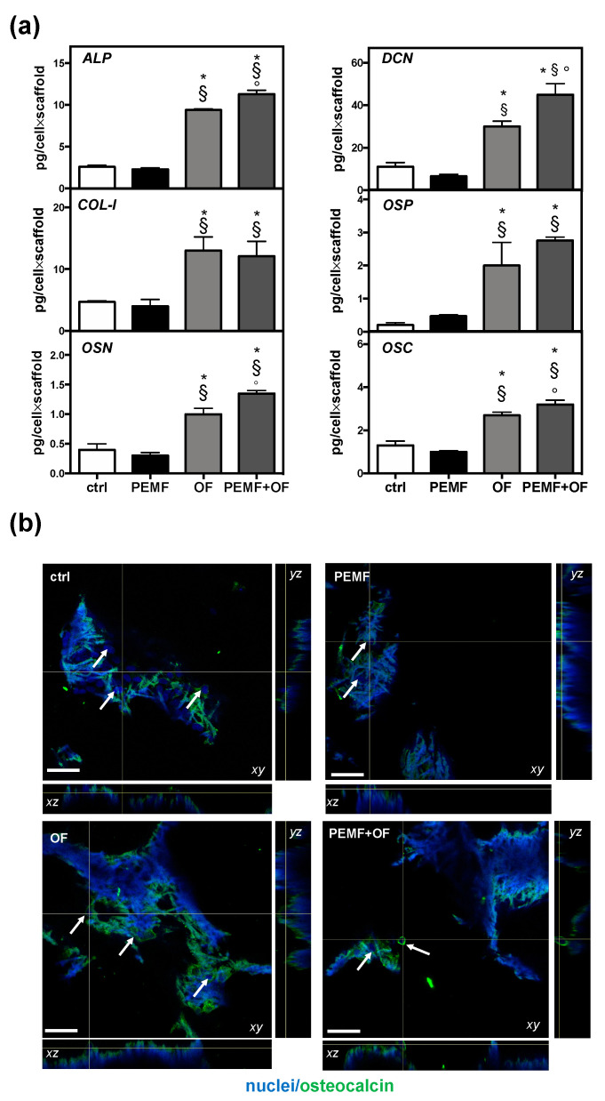
Pulsed electromagnetic field (PEMF) has drawn attention as a potential tool to improve the ability of bone biomaterials to integrate into the surrounding tissue. We investigated the effects of PEMF (frequency, 75 Hz; magnetic induction amplitude, 2 mT; pulse duration, 1.3 ms) on human osteoblast-like cells (SAOS-2) seeded onto wool keratin scaffolds in terms of proliferation, differentiation, and production of the calcified bone extracellular matrix. The wool keratin scaffold offered a 3D porous architecture for cell guesting and nutrient diffusion, suggesting its possible use as a filler to repair bone defects. Here, the combined approach of applying a daily PEMF exposure with additional osteogenic factors stimulated the cells to increase both the deposition of bone-related proteins and calcified matrix onto the wool keratin scaffolds. Also, the presence of SAOS-2 cells, or PEMF, or osteogenic factors did not influence the compression behavior or the resilience of keratin scaffolds in wet conditions. Besides, ageing tests revealed that wool keratin scaffolds were very stable and showed a lower degradation rate compared to commercial collagen sponges. It is for these reasons that this tissue engineering strategy, which improves the osteointegration properties of the wool keratin scaffold, may have a promising application for long term support of bone formation in vivo.

摘要

脉冲电磁场(PEMF)作为一种潜在工具,可提高骨生物材料与周围组织整合的能力,已引起关注。我们研究了PEMF(频率75Hz;磁感应强度2mT;脉冲持续时间1.3ms)对接种在羊毛角蛋白支架上的人成骨样细胞(SAOS-2)在增殖、分化和钙化骨细胞外基质产生方面的影响。羊毛角蛋白支架为细胞寄居和营养物质扩散提供了三维多孔结构,表明其可能用作修复骨缺损的填充物。在此,每日PEMF暴露与额外成骨因子相结合的方法刺激细胞增加骨相关蛋白的沉积以及在羊毛角蛋白支架上的钙化基质。此外,SAOS-2细胞、PEMF或成骨因子的存在并不影响湿态下角蛋白支架的压缩行为或弹性。此外,老化试验表明,与商用胶原海绵相比,羊毛角蛋白支架非常稳定,降解率较低。正是由于这些原因,这种改善羊毛角蛋白支架骨整合特性的组织工程策略可能在体内长期支持骨形成方面具有广阔的应用前景。

https://cdn.ncbi.nlm.nih.gov/pmc/blobs/aa0d/7411850/a489941b75ef/materials-13-03052-g005.jpg

相似文献

1
In Vitro Production of Calcified Bone Matrix onto Wool Keratin Scaffolds via Osteogenic Factors and Electromagnetic Stimulus.通过成骨因子和电磁刺激在羊毛角蛋白支架上体外生成钙化骨基质
Materials (Basel). 2020 Jul 8;13(14):3052. doi: 10.3390/ma13143052.
2
Osteogenic differentiation potential of mesenchymal stem cells cultured on nanofibrous scaffold improved in the presence of pulsed electromagnetic field.在脉冲电磁场存在的情况下,培养在纳米纤维支架上的间充质干细胞的成骨分化潜能得到改善。
J Cell Physiol. 2018 Feb;233(2):1061-1070. doi: 10.1002/jcp.25962. Epub 2017 Jun 6.
3
Wool fibril sponges with perspective biomedical applications.具有潜在生物医学应用前景的羊毛原纤维海绵。
Mater Sci Eng C Mater Biol Appl. 2016 Apr 1;61:42-50. doi: 10.1016/j.msec.2015.11.073. Epub 2015 Dec 2.
4
A comparative analysis of the in vitro effects of pulsed electromagnetic field treatment on osteogenic differentiation of two different mesenchymal cell lineages.脉冲电磁场治疗对两种不同间充质细胞谱系成骨分化的体外效应的比较分析。
Biores Open Access. 2013 Aug;2(4):283-94. doi: 10.1089/biores.2013.0016.
5
Effects of electromagnetic stimulation on calcified matrix production by SAOS-2 cells over a polyurethane porous scaffold.电磁刺激对SAOS-2细胞在聚氨酯多孔支架上产生钙化基质的影响。
Tissue Eng. 2006 Jul;12(7):1985-99. doi: 10.1089/ten.2006.12.1985.
6
The effect of pulsed electromagnetic field exposure on osteoinduction of human mesenchymal stem cells cultured on nano-TiO2 surfaces.脉冲电磁场暴露对纳米 TiO2 表面培养的人骨髓间充质干细胞成骨诱导的影响。
PLoS One. 2018 Jun 14;13(6):e0199046. doi: 10.1371/journal.pone.0199046. eCollection 2018.
7
Fabrication of nanofibrous scaffold using a PLA and hagfish thread keratin composite; its effect on cell adherence, growth, and osteoblast differentiation.采用 PLA 和八目鳗鱼线角蛋白复合材料制备纳米纤维支架;其对细胞黏附、生长和成骨细胞分化的影响。
Biomed Mater. 2013 Aug;8(4):045006. doi: 10.1088/1748-6041/8/4/045006. Epub 2013 Jun 4.
8
Pulsed electromagnetic field may accelerate in vitro endochondral ossification.脉冲电磁场可能会加速体外软骨内成骨。
Bioelectromagnetics. 2015 Jan;36(1):35-44. doi: 10.1002/bem.21882. Epub 2014 Oct 30.
9
Synergism of Electrospun Nanofibers and Pulsed Electromagnetic Field on Osteogenic Differentiation of Induced Pluripotent Stem Cells.静电纺纳米纤维与脉冲电磁场对诱导多能干细胞成骨分化的协同作用。
ASAIO J. 2018 Mar/Apr;64(2):253-260. doi: 10.1097/MAT.0000000000000631.
10
Differentiation of osteoprogenitor cells is induced by high-frequency pulsed electromagnetic fields.高频脉冲电磁场可诱导骨祖细胞分化。
J Craniofac Surg. 2012 Mar;23(2):586-93. doi: 10.1097/SCS.0b013e31824cd6de.

引用本文的文献

1
Injectable bioactive scaffold able to stimulate oral bone regeneration on demand.可按需刺激口腔骨再生的可注射生物活性支架。
J Mater Sci Mater Med. 2025 Apr 8;36(1):31. doi: 10.1007/s10856-025-06879-2.
2
Is Silver Addition to Scaffolds Based on Polycaprolactone Blended with Calcium Phosphates Able to Inhibit and Adhesion and Biofilm Formation?在与磷酸钙混合的聚己内酯基支架中添加银能否抑制细菌黏附和生物膜形成?
Int J Mol Sci. 2024 Feb 28;25(5):2784. doi: 10.3390/ijms25052784.
3
Electron Beam Powder Bed Fusion of Ti-48Al-2Cr-2Nb Open Porous Scaffold for Biomedical Applications: Process Parameters, Adhesion, and Proliferation of NIH-3T3 Cells.

本文引用的文献

1
Keratin - Based materials for biomedical applications.用于生物医学应用的角蛋白基材料。
Bioact Mater. 2020 Apr 16;5(3):496-509. doi: 10.1016/j.bioactmat.2020.04.007. eCollection 2020 Sep.
2
Extracellular matrix-cell interactions: Focus on therapeutic applications.细胞外基质-细胞相互作用:聚焦治疗应用。
Cell Signal. 2020 Feb;66:109487. doi: 10.1016/j.cellsig.2019.109487. Epub 2019 Nov 26.
3
Collagen and non-collagenous proteins molecular crosstalk in the pathophysiology of osteoporosis.胶原蛋白和非胶原蛋白在骨质疏松症病理生理学中的分子对话。
用于生物医学应用的Ti-48Al-2Cr-2Nb开孔多孔支架的电子束粉末床熔融:工艺参数、NIH-3T3细胞的黏附与增殖
3D Print Addit Manuf. 2024 Feb 1;11(1):314-322. doi: 10.1089/3dp.2022.0108. Epub 2024 Feb 15.
4
Effect of Low Copper Doping on the Optical, Cytocompatible, Antibacterial, and SARS-CoV-2 Trapping Properties of Calcium Phosphate Glasses.低铜掺杂对磷酸钙玻璃的光学、细胞相容性、抗菌及新冠病毒捕获性能的影响
ACS Omega. 2023 Nov 1;8(45):42264-42274. doi: 10.1021/acsomega.3c04293. eCollection 2023 Nov 14.
5
Fabrication, Structural Characteristics, and Properties of Sericin-Coated Wool Nonwoven Fabrics.丝胶涂层羊毛非织造布的制备、结构特征和性能。
Int J Mol Sci. 2023 Sep 29;24(19):14750. doi: 10.3390/ijms241914750.
6
Keratose Self-Cross-Linked Wound Dressing for Iron Sequestration in Chronic Wounds.用于慢性伤口铁螯合的角质化自交联伤口敷料。
ACS Omega. 2023 Aug 8;8(33):30118-30128. doi: 10.1021/acsomega.3c02525. eCollection 2023 Aug 22.
7
Human Ovarian Follicular Fluid Mesenchymal Stem Cells Express Osteogenic Markers When Cultured on Bioglass 58S-Coated Titanium Scaffolds.人卵巢卵泡液间充质干细胞在生物玻璃58S涂层钛支架上培养时表达成骨标志物。
Materials (Basel). 2023 May 11;16(10):3676. doi: 10.3390/ma16103676.
8
Harnessing electromagnetic fields to assist bone tissue engineering.利用电磁场辅助骨组织工程。
Stem Cell Res Ther. 2023 Jan 11;14(1):7. doi: 10.1186/s13287-022-03217-z.
9
Long-term stimulation with alternating electric fields modulates the differentiation and mineralization of human pre-osteoblasts.交变电场长期刺激可调节人原代成骨细胞的分化和矿化。
Front Physiol. 2022 Sep 30;13:965181. doi: 10.3389/fphys.2022.965181. eCollection 2022.
10
Haralick's texture analysis to predict cellular proliferation on randomly oriented electrospun nanomaterials.利用哈拉利克纹理分析预测随机取向电纺纳米材料上的细胞增殖
Nanoscale Adv. 2022 Feb 16;4(5):1330-1335. doi: 10.1039/d1na00890k. eCollection 2022 Mar 1.
Cytokine Growth Factor Rev. 2019 Oct;49:59-69. doi: 10.1016/j.cytogfr.2019.09.001. Epub 2019 Sep 13.
4
Permanently hydrophilic, piezoelectric PVDF nanofibrous scaffolds promoting unaided electromechanical stimulation on osteoblasts.具有永久亲水性、压电性的 PVDF 纳米纤维支架促进成骨细胞的自主机电刺激。
Nanoscale. 2019 May 9;11(18):8906-8917. doi: 10.1039/c8nr10384d.
5
Oxidized alginate hydrogels with the GHK peptide enhance cord blood mesenchymal stem cell osteogenesis: A paradigm for metabolomics-based evaluation of biomaterial design.含 GHK 肽的氧化藻酸盐水凝胶增强脐血间充质干细胞成骨作用:基于代谢组学评估生物材料设计的范例。
Acta Biomater. 2019 Apr 1;88:224-240. doi: 10.1016/j.actbio.2019.02.017. Epub 2019 Feb 14.
6
Effects of Electromagnetic Field on Proliferation, Differentiation, and Mineralization of MC3T3 Cells.电磁场对 MC3T3 细胞增殖、分化和矿化的影响。
Tissue Eng Part C Methods. 2019 Feb;25(2):114-125. doi: 10.1089/ten.TEC.2018.0364.
7
Biomaterials and biophysical stimuli for bone regeneration.用于骨再生的生物材料和生物物理刺激。
J Biol Regul Homeost Agents. 2018;32(6 Suppl. 1):41-49.
8
The Use of Pulsed Electromagnetic Fields to Promote Bone Responses to Biomaterials and .使用脉冲电磁场促进骨骼对生物材料的反应以及…… (原文此处不完整)
Int J Biomater. 2018 Sep 3;2018:8935750. doi: 10.1155/2018/8935750. eCollection 2018.
9
The effect of pulsed electromagnetic field exposure on osteoinduction of human mesenchymal stem cells cultured on nano-TiO2 surfaces.脉冲电磁场暴露对纳米 TiO2 表面培养的人骨髓间充质干细胞成骨诱导的影响。
PLoS One. 2018 Jun 14;13(6):e0199046. doi: 10.1371/journal.pone.0199046. eCollection 2018.
10
Pulsed electromagnetic fields increase osteogenetic commitment of MSCs via the mTOR pathway in TNF-α mediated inflammatory conditions: an in-vitro study.脉冲电磁场通过 mTOR 通路增加 TNF-α 介导的炎症条件下间充质干细胞的成骨分化:一项体外研究。
Sci Rep. 2018 Mar 23;8(1):5108. doi: 10.1038/s41598-018-23499-9.