Department of Thoracic Medicine, Royal Adelaide Hospital, Adelaide, SA, Australia.
Adelaide Medical School, University of Adelaide, Adelaide, SA, Australia.
Physiol Rep. 2020 Jul;8(13):e14419. doi: 10.14814/phy2.14419.
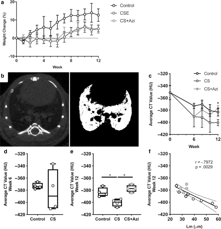
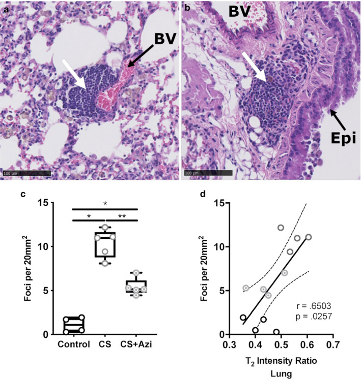
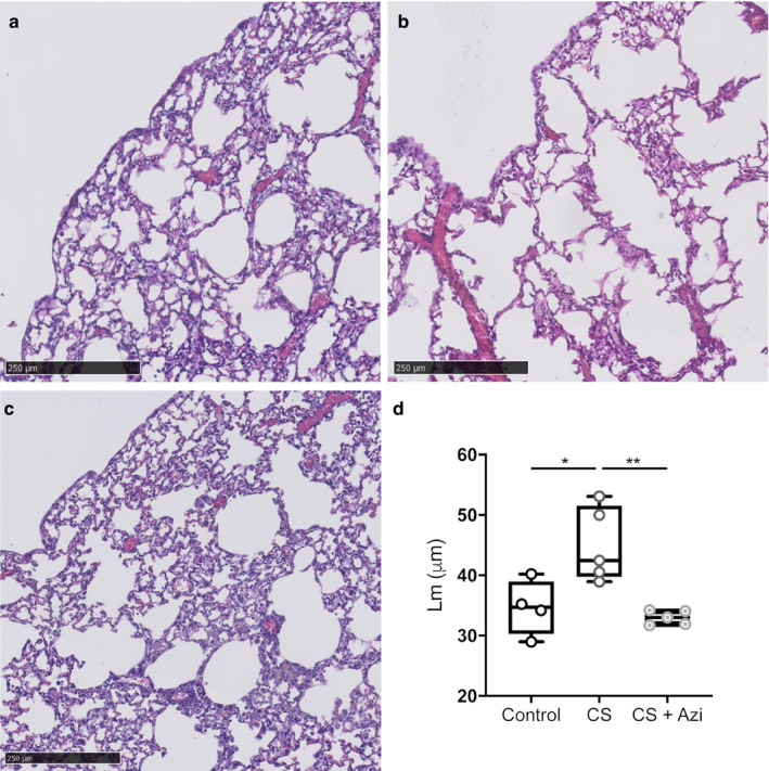
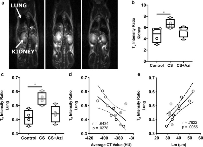
Cigarette smoke (CS)-induced emphysema is an important contributor to chronic obstructive pulmonary disease (COPD). We have shown the efficacy of azithromycin in reducing airway inflammation in COPD and in reducing exacerbations in severe asthma; however, the effects of long-term azithromycin on emphysema development have not been shown. We employed live animal imaging to monitor emphysema-like development and the effects of interventional azithromycin treatment in CS-exposed mice. BALB/c mice (female, 10 weeks; n = 10) were exposed to CS for 1 hr twice daily, 5 days/week, and for 12 weeks (CS). Half were cotreated with low-dose azithromycin during weeks 7-12 (CS + Azi; 0.2 mg kg day ). Microcomputed tomography (CT) and magnetic resonance imaging (MRI) scans were acquired longitudinally. Histological examinations were performed post mortem (mean linear intercept (Lm) and leukocyte infiltration). CS increased median Lm (CS: 42.45 µm versus control: 34.7 µm; p = .0317), this was recovered in CS + Azi mice (33.03 µm). Average CT values were reduced in CS mice (CS: -399.5 Hounsfield units (HU) versus control: -384.9 HU; p = .0286) but not in CS + Azi mice (-377.3 HU). CT values negatively correlated with Lm (r = -.7972; p = .0029) and T -weighted MRI (r = -.6434; p = .0278). MRI also showed significant CS-induced inflammatory changes that were attenuated by azithromycin in the lungs, and positively correlated with Lm (r = .7622; p = .0055) and inflammatory foci counts (r = .6503; p = .0257). Monitoring of emphysema development is possible via micro-CT and MRI. Interventional azithromycin treatment in CS-exposed mice attenuated the development of pulmonary emphysema-like changes.
香烟烟雾(CS)引起的肺气肿是慢性阻塞性肺疾病(COPD)的重要原因。我们已经证明了阿奇霉素在减轻 COPD 气道炎症和减少重度哮喘恶化方面的疗效;然而,长期使用阿奇霉素对肺气肿发展的影响尚未得到证实。我们采用活体动物成像技术监测 CS 暴露小鼠的肺气肿样发展和干预性阿奇霉素治疗的效果。BALB/c 小鼠(雌性,10 周;n=10)每天暴露于 CS 两次,每次 1 小时,每周 5 天,共 12 周(CS)。其中一半在第 7-12 周进行低剂量阿奇霉素联合治疗(CS+Azi;0.2mg/kg/天)。纵向采集微计算机断层扫描(CT)和磁共振成像(MRI)扫描。死后进行组织学检查(平均线性截距(Lm)和白细胞浸润)。CS 增加了中位数 Lm(CS:42.45µm 与对照:34.7µm;p=0.0317),CS+Azi 组小鼠的 Lm 恢复到正常水平(33.03µm)。CS 组小鼠的平均 CT 值降低(CS:-399.5 亨斯菲尔德单位(HU)与对照:-384.9 HU;p=0.0286),但 CS+Azi 组小鼠的 CT 值未降低(-377.3 HU)。CT 值与 Lm 呈负相关(r=-0.7972;p=0.0029)和 T 加权 MRI(r=-0.6434;p=0.0278)。MRI 还显示 CS 引起的肺部炎症变化显著,阿奇霉素治疗可减轻这些变化,与 Lm(r=0.7622;p=0.0055)和炎症灶计数(r=0.6503;p=0.0257)呈正相关。通过微 CT 和 MRI 可以监测肺气肿的发展。CS 暴露小鼠的干预性阿奇霉素治疗减轻了肺肺气肿样改变的发展。