Fu Hongjun, Liu Bin, Li Liangping, Lemere Cynthia A
Department of Neuroscience, Chronic Brain Injury, Discovery Themes, The Ohio State University, Columbus, OH, USA; Ann Romney Center for Neurologic Diseases, Brigham and Women's Hospital, Harvard Medical School, Boston, MA, USA.
Ann Romney Center for Neurologic Diseases, Brigham and Women's Hospital, Harvard Medical School, Boston, MA, USA.
Neuroscience. 2020 Sep 1;443:30-43. doi: 10.1016/j.neuroscience.2020.07.020. Epub 2020 Jul 19.
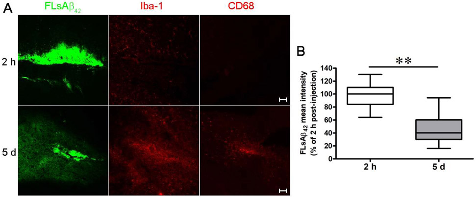
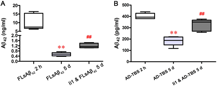
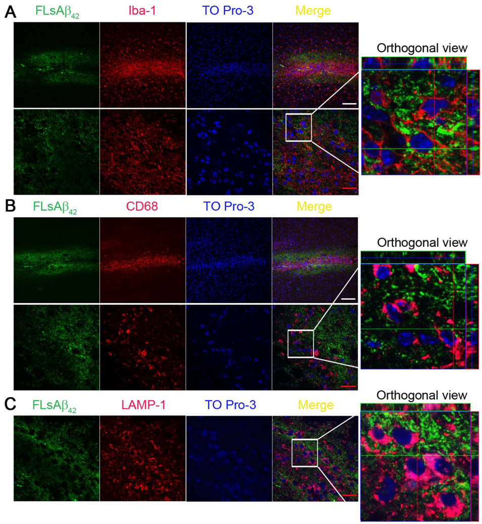
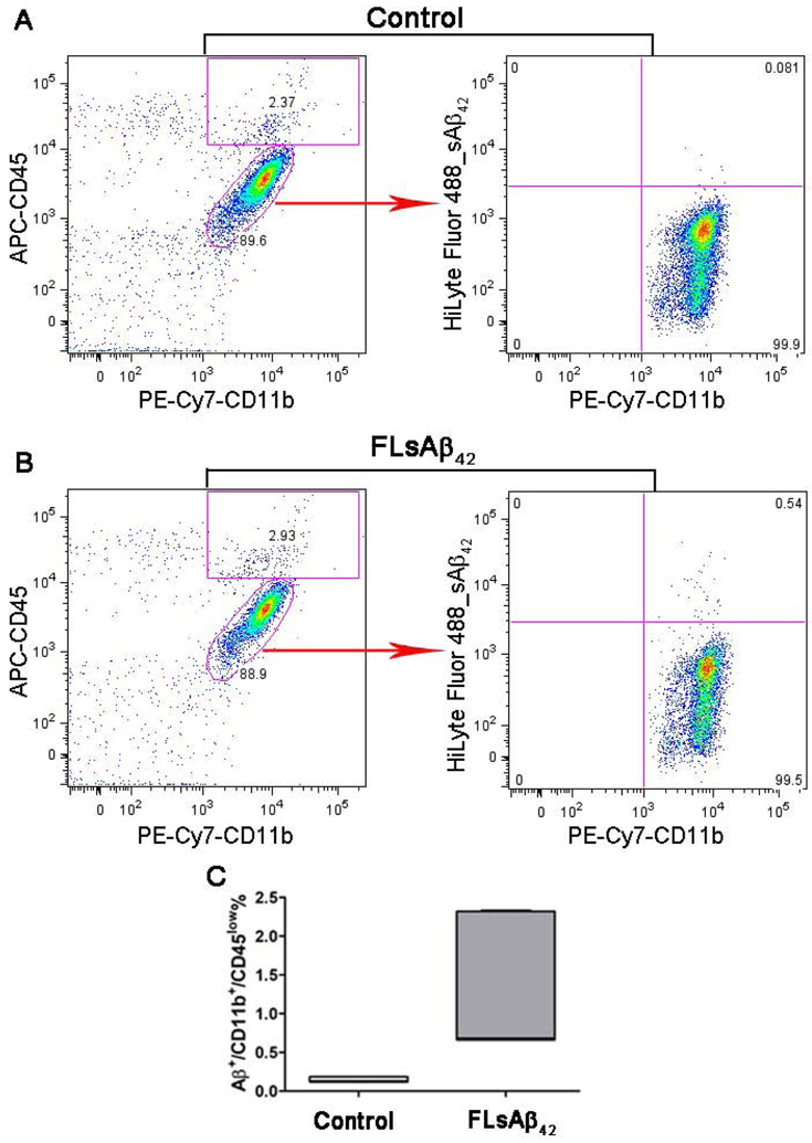
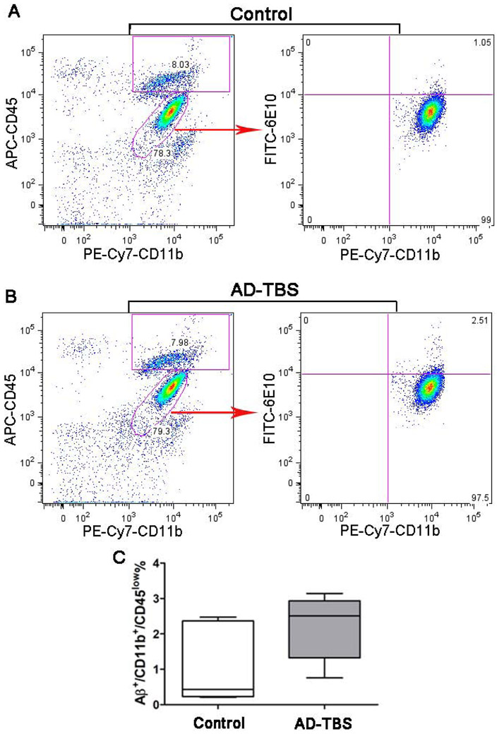
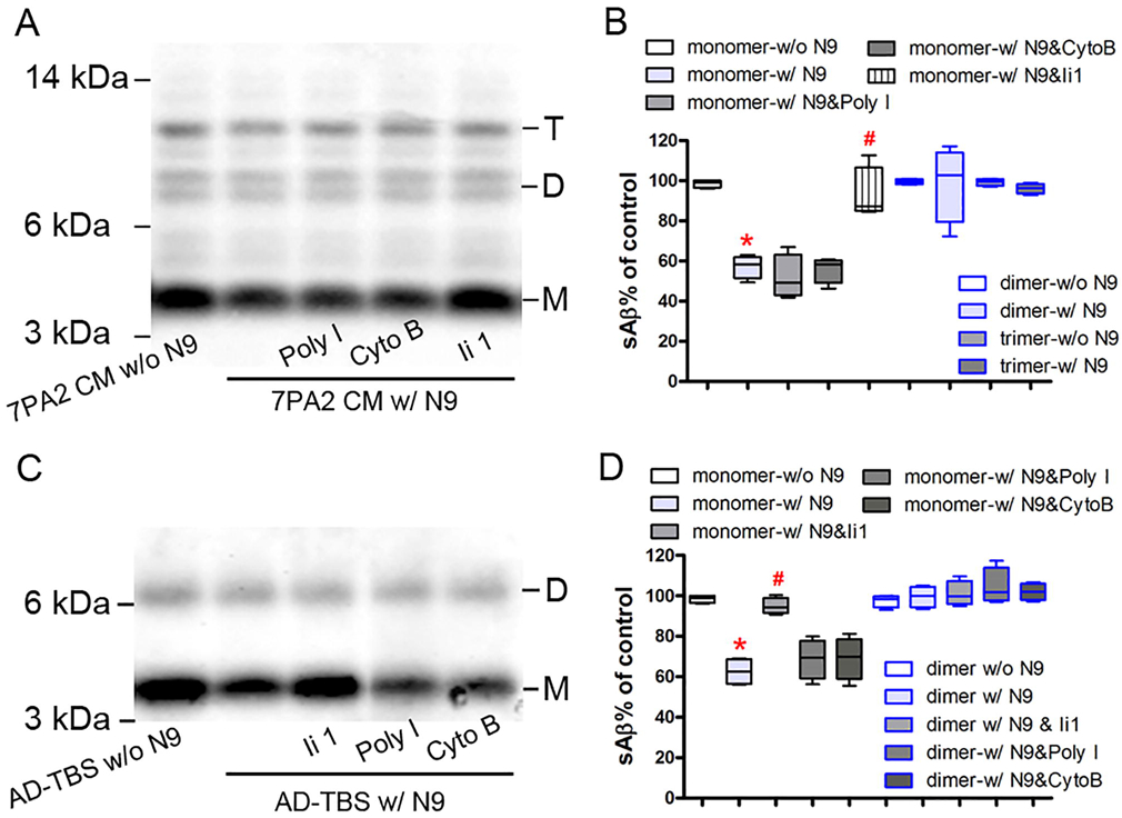
Microglia play important roles in the pathogenesis of Alzheimer's disease (AD), in part, by affecting the clearance of amyloid-β (Aβ) peptides. Most studies, however, used synthetic soluble Aβ (sAβ) at higher concentrations. The exact mechanisms underlying microglia-mediated clearance of physiological sAβ at very low concentrations remain unclear. Here we reported that there were much more Iba-1- and CD68-positive microglia and significantly less sAβ left in the brain of adult mice 5 days after the surgery of sAβ microinjection compared to 2 h after the surgery (p < 0.05). However, very few Iba-1- and CD68-positive microglia co-localized with microinjected fluorescently labeled sAβ (FLsAβ) 5 days after the surgery. Also, there was no co-localization of FLsAβ with a lysosomal marker (LAMP-1) 5 days after the surgery. There was no significant difference in the percentage of Aβ/PE-CD11b/APC-CD45 microglia between the control group and the group microinjected with TBS-soluble Aβ extracted from the brains of AD patients (p > 0.05). The degradation of physiological sAβ was prevented by a highly selective insulin-degrading enzyme inhibitor (Ii1) but not by a phagocytosis inhibitor (polyinosinic acid) or pinocytosis inhibitor (cytochalasin B) in vitro. Furthermore, the reduction of synthetic and physiological sAβ in the brain was partially prevented by the co-injection of Ii1 in vivo (p < 0.05). Our results demonstrate that microglia do not take up synthetic or physiological sAβ, but partially degrade it via the secretion of insulin-degrading enzyme, which will be beneficial for understanding how sAβ is removed from the brain by microglia.
小胶质细胞在阿尔茨海默病(AD)的发病机制中发挥着重要作用,部分原因是通过影响淀粉样β(Aβ)肽的清除。然而,大多数研究使用的是较高浓度的合成可溶性Aβ(sAβ)。小胶质细胞在极低浓度下介导生理性sAβ清除的确切机制仍不清楚。在此,我们报告,与注射后2小时相比,成年小鼠在sAβ微量注射手术后5天,脑内Iba-1和CD68阳性小胶质细胞更多,剩余的sAβ显著减少(p<0.05)。然而,手术后5天,很少有Iba-1和CD68阳性小胶质细胞与微量注射的荧光标记sAβ(FLsAβ)共定位。此外,手术后5天,FLsAβ与溶酶体标记物(LAMP-1)没有共定位。对照组与注射从AD患者脑中提取的TBS可溶性Aβ的组之间,Aβ/PE-CD11b/APC-CD45小胶质细胞的百分比没有显著差异(p>0.05)。体外实验中,高选择性胰岛素降解酶抑制剂(Ii1)可阻止生理性sAβ的降解,但吞噬抑制剂(聚肌苷酸)或胞饮抑制剂(细胞松弛素B)则不能。此外,体内共注射Ii1可部分阻止脑内合成和生理性sAβ的减少(p<0.05)。我们的结果表明,小胶质细胞不摄取合成或生理性sAβ,而是通过分泌胰岛素降解酶部分降解它,这将有助于理解小胶质细胞如何从脑中清除sAβ。