Pan Xia, Zhang Kai, Shen Cheng, Wang Xi, Wang Long, Huang Ya-Yi
Department of Anesthesiology, Renmin Hospital of Wuhan University, Wuhan, Hubei 430060, China.
Department of Cardiology, The Affiliated Hospital of Guizhou Medical University, Guiyang, Guizhou 550001, China.
Chin Med J (Engl). 2020 Aug 5;133(15):1786-1797. doi: 10.1097/CM9.0000000000000814.
Cardiac remodeling after acute myocardial infarction (AMI) is an important process. The present study aimed to assess the protective effects of astaxanthin (ASX) on cardiac remodeling after AMI.
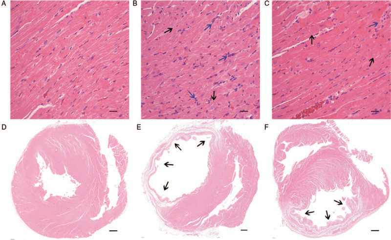
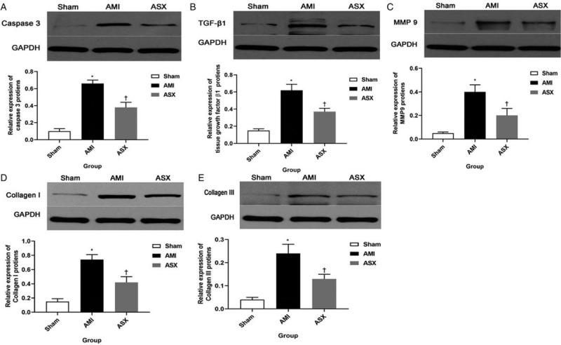
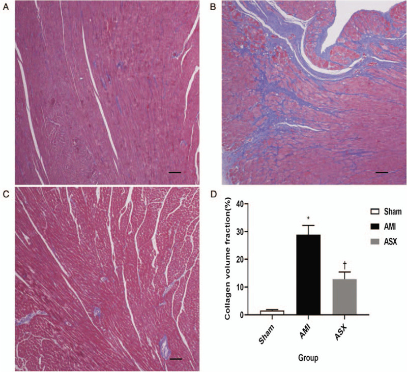
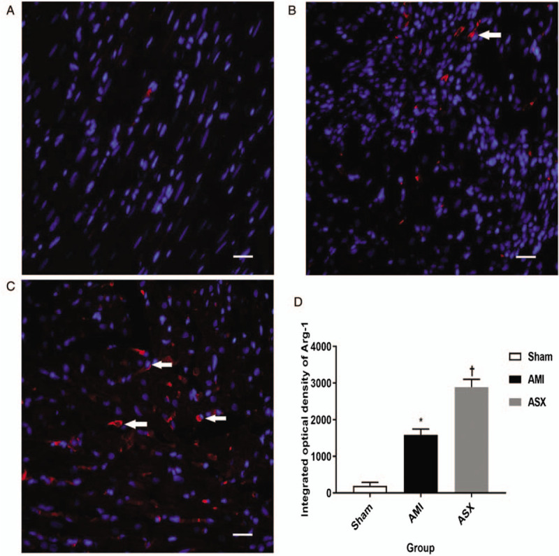
The study was conducted between April and September 2018. To create a rat AMI model, rats were anesthetized, and the left anterior descending coronary artery was ligated. The rats in the ASX group received 10 mg·kg·day ASX by gavage for 28 days. On the 1st day after AMI, but before ASX administration, six rats from each group were sacrificed to evaluate changes in the heart function and peripheral blood (PB) levels of inflammatory factors. On the 7th day after AMI, eight rats from each group were sacrificed to evaluate the PB levels of inflammatory factors and the M2 macrophage count using both immunofluorescence (IF) and flow cytometry (FC). The remaining rats were observed for 28 days. Cardiac function was examined using echocardiography. The inflammatory factors, namely, tumor necrosis factor-α (TNF-α), interleukin-1β (IL-1β), and IL-10, were assessed using enzyme-linked immunosorbent assay. The heart weight/body weight (BW), and lung weight (LW)/BW ratios were calculated, and myocardial fibrosis in the form of collagen volume fraction was measured using Masson trichrome staining. Hematoxylin and eosin (H&E) staining was used to determine the myocardial infarct size (MIS), and TdT-mediated dUTP nick-end labeling staining was used to analyze the myocardial apoptosis index. The levels of apoptosis-related protein, type I/III collagen, transforming growth factor β1 (TGF-β1), metalloproteinase 9 (MMP9), and caspase 3 were assessed by Western blotting. Unpaired t-test, one-way analysis of variance, and non-parametric Mann-Whitney test were used to analyze the data.
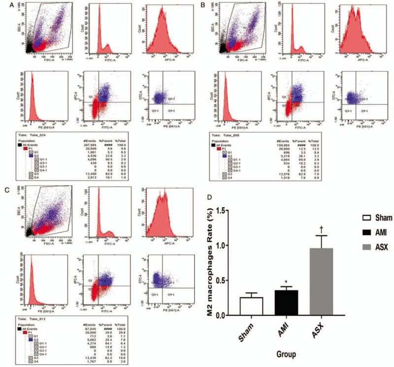
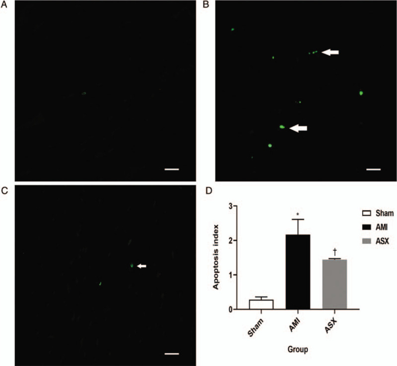
On day 1, cardiac function was worse in the ASX group than in the sham group (left ventricular end-systolic diameter [LVIDs]: 0.72 ± 0.08 vs. 0.22 ± 0.06 cm, t = -11.38; left ventricular end-diastolic diameter [LVIDd]: 0.89 ± 0.09 vs. 0.48 ± 0.05 cm, t = -9.42; end-systolic volume [ESV]: 0.80 [0.62, 0.94] vs. 0.04 [0.03, 0.05] mL, Z = -2.89; end-diastolic volume [EDV]: 1.39 [1.03, 1.49] vs. 0.28 [0.22, 0.32] mL, Z = -2.88; ejection fraction [EF]: 0.40 ± 0.04 vs. 0.86 ± 0.05, t = 10.00; left ventricular fractional shortening [FS] rate: 0.19 [0.18, 0.20] %FS vs. 0.51 [0.44, 0.58] %FS, Z = -2.88, all P < 0.01; n = 6). The levels of inflammatory factors significantly increased (TNF-α: 197.60 [133.89, 237.94] vs. 50.48 [47.21 57.10] pg/mL, Z = -2.88; IL-1β: 175.23 [160.74, 215.09] vs. 17.78 [16.83, 19.56] pg/mL, Z = -2.88; IL-10: 67.64 [58.90, 71.46] vs. 12.33 [11.64, 13.98] pg/mL, Z = -2.88, all P < 0.01; n = 6). On day 7, the levels of TNF-α and IL-1β were markedly lower in the ASX group than in the AMI group (TNF-α: 71.70 [68.60, 76.00] vs. 118.07 [106.92, 169.08] pg/mL, F = 42.64; IL-1β: 59.90 [50.83, 73.78] vs. 151.60 [108.4, 198.36] pg/mL, F = 44.35, all P < 0.01, n = 8). Conversely, IL-10 levels significantly increased (141.84 [118.98, 158.36] vs. 52.96 [42.68, 74.52] pg/mL, F = 126.67, P < 0.01, n = 8). The M2 macrophage count significantly increased (2891.42 ± 211.29 vs. 1583.38 ± 162.22, F = 274.35, P < 0.01 by immunofluorescence test; 0.96 ± 0.18 vs. 0.36 ± 0.05, F = 46.24, P < 0.05 by flowcytometry test). On day 28, cardiac function was better in the ASX group than in the AMI group (LVIDs: 0.50 [0.41, 0.56] vs. 0.64 [0.56, 0.74] cm, Z = -3.60; LVIDd: 0.70 [0.60, 0.76] vs. 0.80 [0.74 0.88] cm, Z = -2.96; ESV: 0.24 [0.18, 0.45] vs. 0.58 [0.44, 0.89] mL, Z = -3.62; EDV: 0.76 [0.44, 1.04] vs. 1.25 [0.82, 1.46] mL, Z = -2.54; EF: 0.60 ± 0.08 vs. 0.50 ± 0.12, F = 160.48; %FS: 0.29 [0.24, 0.31] vs. 0.20 [0.17, 0.21], Z = -4.43, all P < 0.01; n = 16). The MIS and LW/BW ratio were markedly lower in the ASX group than in the AMI group (myocardial infarct size: 32.50 ± 1.37 vs. 50.90 ± 1.73, t = 23.63, P < 0.01, n = 8; LW/BW: 1.81 ± 0.15 vs. 2.17 ± 0.37, t = 3.66, P = 0.01, n = 16). The CVF was significantly lower in the ASX group than in the AMI group: 12.88 ± 2.53 vs. 28.92 ± 3.31, t = 10.89, P < 0.01, n = 8. The expression of caspase 3, TGF-β1, MMP9, and type I/III collagen was lower in the ASX group than in the AMI group (caspase 3: 0.38 ± 0.06 vs. 0.66 ± 0.04, t = 8.28; TGF-β1: 0.37 ± 0.04 vs. 0.62 ± 0.07, t = 6.39; MMP9: 0.20 ± 0.06 vs. 0.40 ± 0.06, t = 4.62; type I collagen: 0.42 ± 0.09 vs. 0.74 ± 0.07, t = 5.73; type III collagen: 0.13 ± 0.02 vs. 0.74 ± 0.07, t = 4.32, all P < 0.01; n = 4).
ASX treatment after AMI may promote M2 macrophages and effectively attenuate cardiac remodeling by inhibiting inflammation and reducing myocardial fibrosis.
急性心肌梗死(AMI)后的心脏重塑是一个重要过程。本研究旨在评估虾青素(ASX)对AMI后心脏重塑的保护作用。
本研究于2018年4月至9月进行。为建立大鼠AMI模型,将大鼠麻醉后结扎左冠状动脉前降支。ASX组大鼠通过灌胃给予10 mg·kg·天的ASX,持续28天。在AMI后第1天,但在给予ASX之前,每组处死6只大鼠,以评估心脏功能和外周血(PB)中炎症因子水平的变化。在AMI后第7天,每组处死8只大鼠,通过免疫荧光(IF)和流式细胞术(FC)评估PB中炎症因子水平和M2巨噬细胞计数。其余大鼠观察28天。使用超声心动图检查心脏功能。使用酶联免疫吸附测定法评估炎症因子,即肿瘤坏死因子-α(TNF-α)、白细胞介素-1β(IL-1β)和IL-10。计算心脏重量/体重(BW)和肺重量(LW)/BW比值,并使用Masson三色染色法测量以胶原体积分数形式存在的心肌纤维化。使用苏木精和伊红(H&E)染色确定心肌梗死面积(MIS),并使用TdT介导的dUTP缺口末端标记染色分析心肌凋亡指数。通过蛋白质印迹法评估凋亡相关蛋白、I/III型胶原、转化生长因子β1(TGF-β1)、金属蛋白酶9(MMP9)和半胱天冬酶3的水平。使用非配对t检验、单因素方差分析和非参数Mann-Whitney检验分析数据。
在第1天,ASX组的心脏功能比假手术组差(左心室收缩末期直径[LVIDs]:0.72±0.08 vs. 0.22±0.06 cm,t = -11.38;左心室舒张末期直径[LVIDd]:0.89±0.09 vs. 0.48±0.05 cm,t = -9.42;收缩末期容积[ESV]:0.80[0.62, 0.94] vs. 0.04[0.03, 0.05] mL,Z = -2.89;舒张末期容积[EDV]:1.39[1.03, 1.49] vs. 0.28[0.22, 0.32] mL,Z = -2.88;射血分数[EF]:0.40±0.04 vs. 0.86±0.05,t = 10.00;左心室短轴缩短率[FS]:0.19[0.18, 0.20]%FS vs. 0.51[0.44, 0.58]%FS,Z = -2.88,所有P < 0.01;n = 6)。炎症因子水平显著升高(TNF-α:197.60[133.89, 237.94] vs. 50.48[47.21, 57.10] pg/mL,Z = -2.88;IL-1β:175.23[160.74, 215.09] vs. 17.78[16.83, 19.56] pg/mL,Z = -2.88;IL-10:67.64[58.90, 71.46] vs. 12.33[11.64, 13.98] pg/mL,Z = -2.88,所有P < 0.01;n = 6)。在第7天,ASX组的TNF-α和IL-1β水平明显低于AMI组(TNF-α:71.70[68.60, 76.00] vs. 118.07[106.92, 169.08] pg/mL,F = 42.64;IL-1β:59.90[50.83, 73.78] vs. 151.60[108.4, 198.36] pg/mL,F = 44.35,所有P < 0.01,n = 8)。相反,IL-10水平显著升高(141.84[118.98, 158.36] vs. 52.96[42.68, 74.52] pg/mL,F = 126.67,P < 0.01,n = 8)。M2巨噬细胞计数显著增加(免疫荧光试验:2,891.42±211.29 vs. 1,583.38±162.22,F = 274.35,P < 0.01;流式细胞术试验:0.96±0.18 vs. 0.36±0.05,F = 46.24,P < 0.05)。在第28天,ASX组的心脏功能比AMI组好(LVIDs:0.50[0.41, 0.56] vs. 0.64[0.56, 0.74] cm,Z = -3.60;LVIDd:0.70[0.60, 0.76] vs. 0.80[0.74, 0.88] cm,Z = -2.96;ESV:0.24[0.18, 0.45] vs. 0.58[0.44, 0.89] mL,Z = -3.62;EDV:0.76[0.44, 1.04] vs. 1.25[0.82, 1.46] mL,Z = -2.54;EF:0.60±0.08 vs. 0.50±0.12,F = 160.48;%FS:0.29[0.24, 0.31] vs. 0.20[0.17, 0.21],Z = -4.43,所有P < 0.01;n = 16)。ASX组的MIS和LW/BW比值明显低于AMI组(心肌梗死面积:32.50±1.37 vs. 50.90±1.73,t = 23.63,P < 0.01,n = 8;LW/BW:1.81±0.15 vs. 2.17±0.37,t = 3.66,P = 0.01,n = 16)。ASX组的CVF明显低于AMI组:12.88±2.53 vs. 28.92±3.