Laboratory of Cell Signalling, Institute of Microbiology of the Czech Academy of Sciences, 142 00 Prague, Czech Republic.
Biomolecules. 2020 Jul 22;10(8):1089. doi: 10.3390/biom10081089.
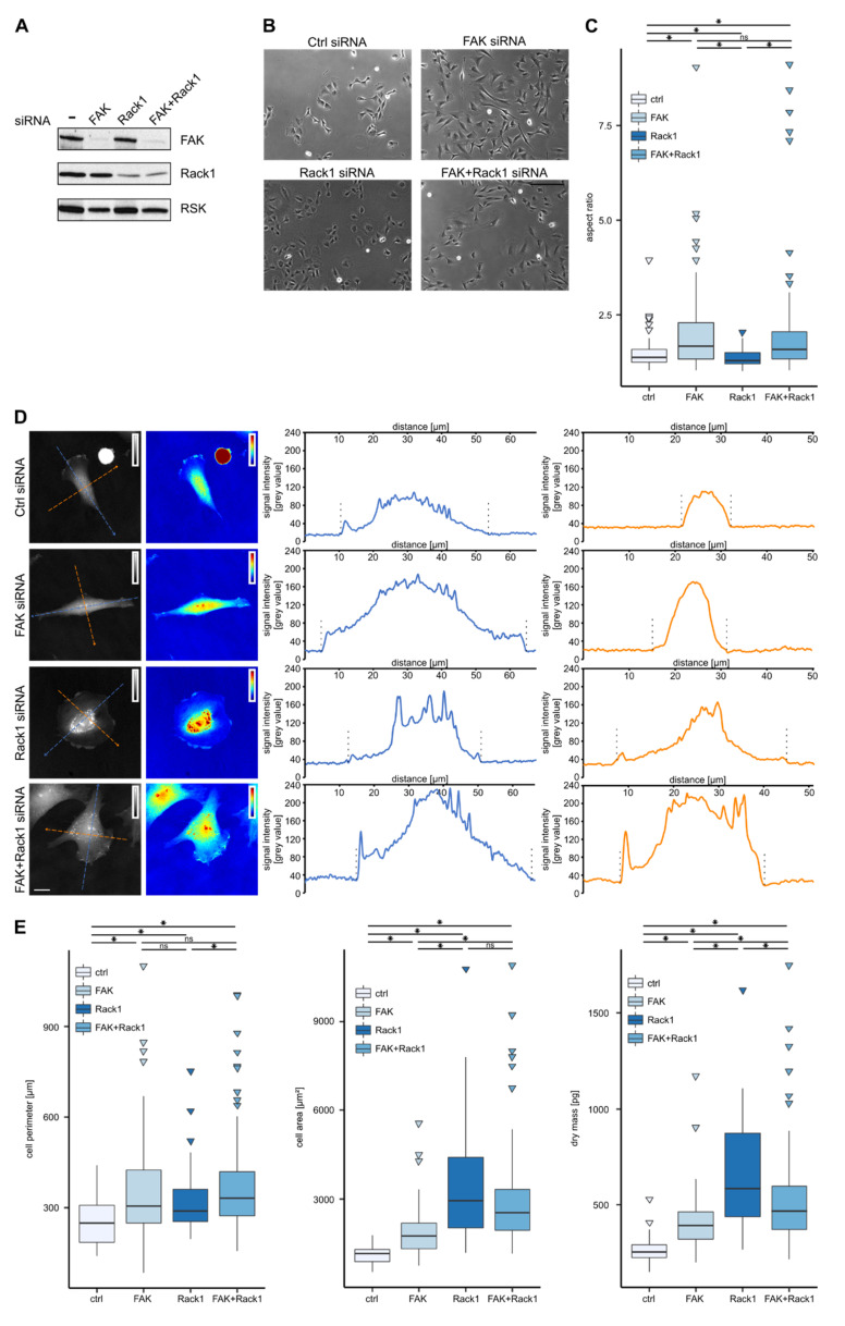
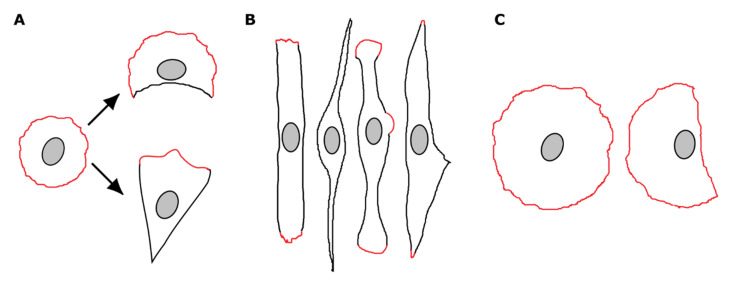
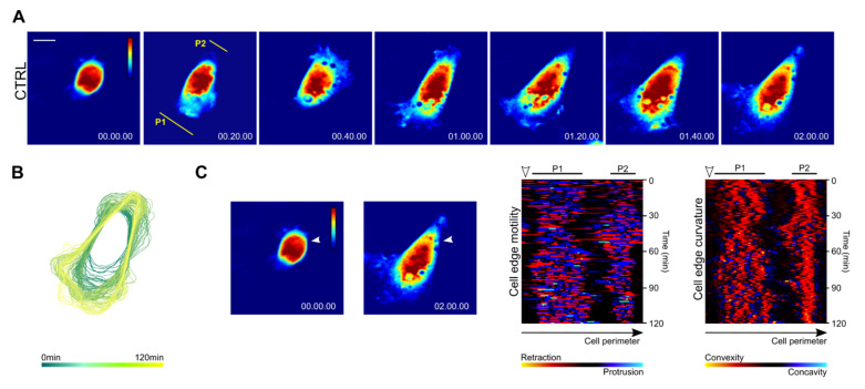
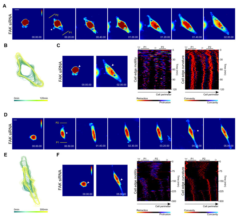
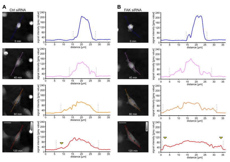
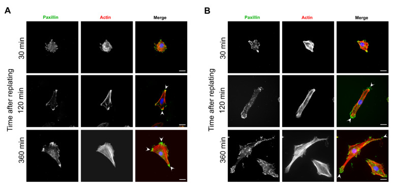
Cells attaching to the extracellular matrix spontaneously acquire front-rear polarity. This self-organization process comprises spatial activation of polarity signaling networks and the establishment of a protruding cell front and a non-protruding cell rear. Cell polarization also involves the reorganization of cell mass, notably the nucleus that is positioned at the cell rear. It remains unclear, however, how these processes are regulated. Here, using coherence-controlled holographic microscopy (CCHM) for non-invasive live-cell quantitative phase imaging (QPI), we examined the role of the focal adhesion kinase (FAK) and its interacting partner Rack1 in dry mass distribution in spreading Rat2 fibroblasts. We found that FAK-depleted cells adopt an elongated, bipolar phenotype with a high central body mass that gradually decreases toward the ends of the elongated processes. Further characterization of spreading cells showed that FAK-depleted cells are incapable of forming a stable rear; rather, they form two distally positioned protruding regions. Continuous protrusions at opposite sides results in an elongated cell shape. In contrast, Rack1-depleted cells are round and large with the cell mass sharply dropping from the nuclear area towards the basal side. We propose that FAK and Rack1 act differently yet coordinately to establish front-rear polarity in spreading cells.
细胞自发地附着到细胞外基质上,从而获得前后极性。这个自我组织过程包括极性信号网络的空间激活,以及突出的细胞前端和非突出的细胞后端的建立。细胞极化还涉及细胞质量的重新组织,特别是细胞核位于细胞后端。然而,这些过程是如何被调控的,目前还不清楚。在这里,我们使用相干控制全息显微镜(CCHM)对非侵入性活细胞定量相衬成像(QPI)进行研究,以检查粘着斑激酶(FAK)及其相互作用伙伴Rack1 在铺展 Rat2 成纤维细胞中干性物质分布中的作用。我们发现,FAK 耗尽的细胞呈现出伸长的双极表型,其中央体质量很高,逐渐向伸长过程的末端降低。对铺展细胞的进一步特征分析表明,FAK 耗尽的细胞无法形成稳定的后端;相反,它们形成两个位于远端的突出区域。在相反侧的连续突出导致细胞呈伸长形状。相比之下,Rack1 耗尽的细胞呈圆形且较大,细胞质量从核区域急剧下降到基底侧。我们提出,FAK 和 Rack1 以不同但协调的方式作用,在铺展细胞中建立前后极性。