Department of Clinical and Movement Neurosciences, UCL Institute of Neurology and The National Hospital for Neurology and Neurosurgery, Queen Square, London, UK.
Nuffield Department of Clinical Neurosciences, John Radcliffe Hospital, Oxford, UK; Wellcome Centre for Human Neuroimaging, UCL Institute of Neurology, 12 Queen Square, London, UK.
Neuroimage. 2020 Nov 1;221:117184. doi: 10.1016/j.neuroimage.2020.117184. Epub 2020 Jul 22.
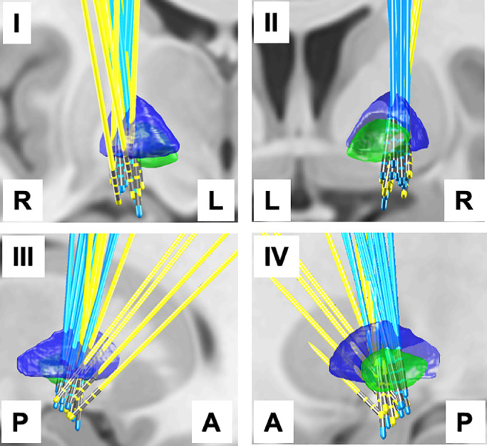
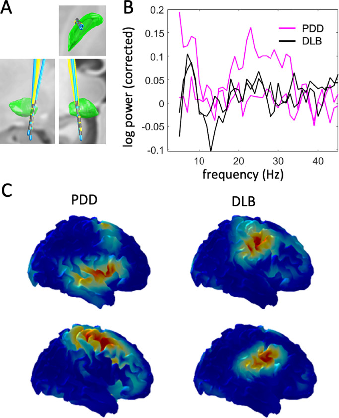
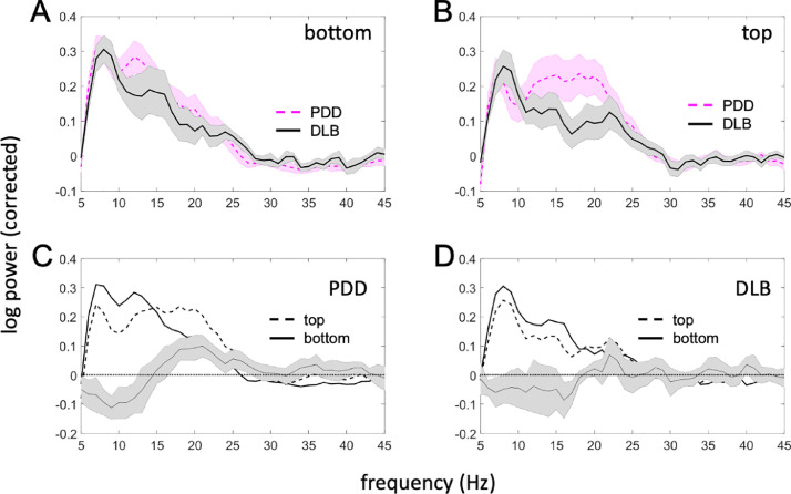
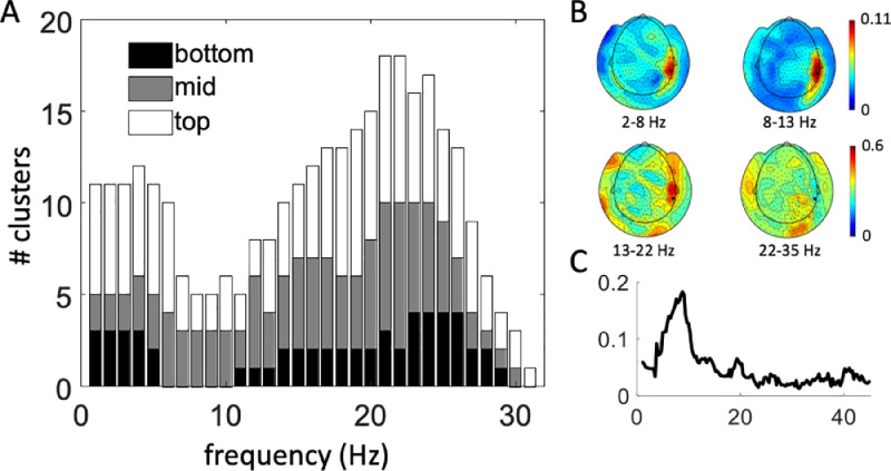
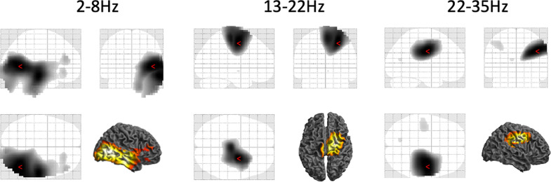
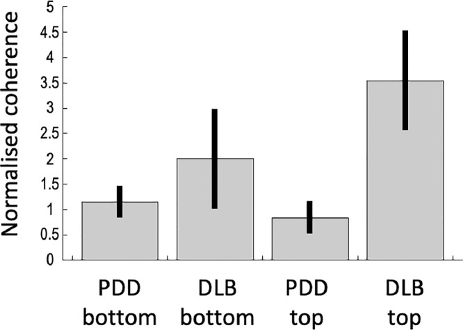
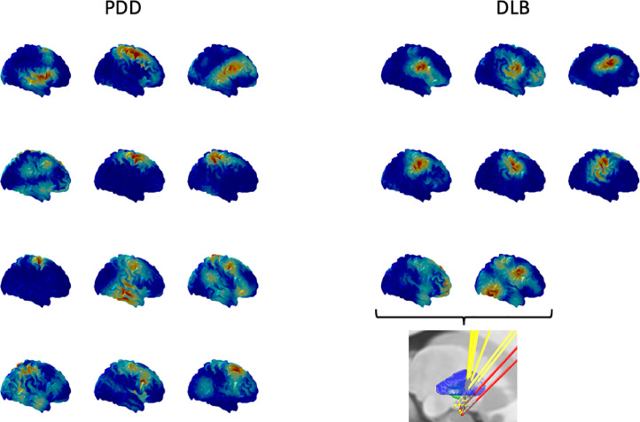
Parkinson's disease dementia (PDD) and dementia with Lewy bodies (DLB) are two related diseases which can be difficult to distinguish. There is no objective biomarker which can reliably differentiate between them. The synergistic combination of electrophysiological and neuroimaging approaches is a powerful method for interrogation of functional brain networks in vivo. We recorded bilateral local field potentials (LFPs) from the nucleus basalis of Meynert (NBM) and the internal globus pallidus (GPi) with simultaneous cortical magnetoencephalography (MEG) in six PDD and five DLB patients undergoing surgery for deep brain stimulation (DBS) to look for differences in underlying resting-state network pathophysiology. In both patient groups we observed spectral peaks in the theta (2-8 Hz) band in both the NBM and the GPi. Furthermore, both the NBM and the GPi exhibited similar spatial and spectral patterns of coupling with the cortex in the two disease states. Specifically, we report two distinct coherent networks between the NBM/GPi and cortical regions: (1) a theta band (2-8 Hz) network linking the NBM/GPi to temporal cortical regions, and (2) a beta band (13-22 Hz) network coupling the NBM/GPi to sensorimotor areas. We also found differences between the two disease groups: oscillatory power in the low beta (13-22Hz) band was significantly higher in the globus pallidus in PDD patients compared to DLB, and coherence in the high beta (22-35Hz) band between the globus pallidus and lateral sensorimotor cortex was significantly higher in DLB patients compared to PDD. Overall, our findings reveal coherent networks of the NBM/GPi region that are common to both DLB and PDD. Although the neurophysiological differences between the two conditions in this study are confounded by systematic differences in DBS lead trajectories and motor symptom severity, they lend support to the hypothesis that DLB and PDD, though closely related, are distinguishable from a neurophysiological perspective.
帕金森病痴呆(PDD)和路易体痴呆(DLB)是两种相关疾病,难以区分。目前尚无可靠的客观生物标志物可将两者区分开来。电生理和神经影像学方法的协同组合是研究活体功能大脑网络的有力方法。我们在六名 PDD 和五名 DLB 患者中记录了双侧基底节核(NBM)和内苍白球(GPi)的局部场电位(LFP),并同时进行皮层脑磁图(MEG)记录,这些患者正在接受深部脑刺激(DBS)手术,以寻找潜在静息态网络病理生理学的差异。在这两个患者组中,我们在 NBM 和 GPi 中均观察到了θ(2-8 Hz)频段的频谱峰值。此外,在两种疾病状态下,NBM 和 GPi 都表现出与皮层相似的空间和频谱耦合模式。具体来说,我们报告了 NBM/GPi 和皮层区域之间存在两个不同的相干网络:(1)一个θ频段(2-8 Hz)网络,将 NBM/GPi 与颞叶皮层区域连接起来;(2)一个β频段(13-22 Hz)网络,将 NBM/GPi 与感觉运动区域耦合。我们还发现了两个疾病组之间的差异:与 DLB 相比,PDD 患者的苍白球低β(13-22 Hz)频段的振荡功率明显更高,而苍白球与外侧感觉运动皮层之间的高β(22-35 Hz)频段的相干性在 DLB 患者中明显更高。总的来说,我们的研究结果揭示了 NBM/GPi 区域的相干网络,这些网络在 DLB 和 PDD 中都是共同的。尽管本研究中两种情况之间的神经生理学差异受到 DBS 导联轨迹和运动症状严重程度的系统差异的影响,但这些差异支持了这样一种假设,即 DLB 和 PDD 虽然密切相关,但从神经生理学的角度来看是可以区分的。