Department of Radiation Medicine and Applied Sciences, University of California San Diego, La Jolla, California.
Department of Radiation Medicine and Applied Sciences, University of California San Diego, La Jolla, California; Center for Multimodal Imaging and Genetics, University of California San Diego, La Jolla, California.
Int J Radiat Oncol Biol Phys. 2020 Dec 1;108(5):1218-1228. doi: 10.1016/j.ijrobp.2020.07.032. Epub 2020 Jul 23.
Our purpose was to investigate the association between imaging biomarkers of radiation-induced white matter (WM) injury within perisylvian regions and longitudinal language decline in patients with brain tumors.
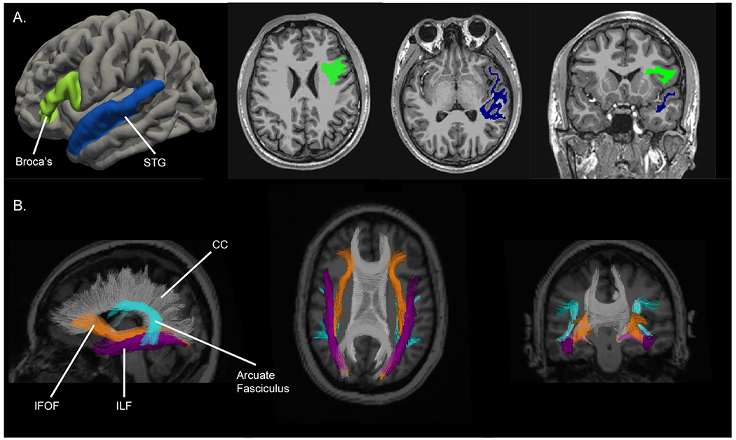
Patients with primary brain tumors (n = 44) on a prospective trial underwent brain magnetic resonance imaging, diffusion-weighted imaging, and language assessments of naming (Boston Naming Test [BNT]) and fluency (Delis-Kaplan Executive Function System Category Fluency [DKEFS-CF]) at baseline and 3, 6, and 12 months after fractionated radiation therapy (RT). Reliable change indices of language function (0-6 months), accounting for practice effects (RCI-PE), evaluated decline. Bilateral perisylvian WM regions (superficial WM subadjacent to Broca's area and the superior temporal gyrus [STG], inferior longitudinal fasciculus [ILF], inferior fronto-occipital fasciculus [IFOF], and arcuate fasciculus) were autosegmented. We quantified volume and diffusion measures of WM microstructure: fractional anisotropy (FA; lower values indicate disruption) and mean diffusivity (MD; higher values indicate injury). Linear mixed-effects models assessed mean dose as predictor of imaging biomarker change and imaging biomarkers as longitudinal predictors of language scores.
DKEFS-CF scores declined at 6 months post-RT (RCI-PE, -0.483; P = .01), whereas BNT scores improved (RCI-PE, 0.262; P = .04). Higher mean dose to left and right regions was predictive of decreased volume (left-STG, P = .02; right-ILF and IFOF, P = .03), decreased FA (left-WM tracts, all P < .01; right-STG and IFOF, P < .02), and increased MD of left-WM tracts (all P < .03). Volume loss within left-Broca's area (P = .01), left-ILF (P = .01), left-IFOF (P = .01), and left-arcuate fasciculus (P = .04) was associated with lower BNT scores. Lower FA correlated with poorer DKEFS-CF and BNT scores within left-ILF (P = .02, not significant), left-IFOF (P = .02, .04), and left-arcuate fasciculus (P = .01, .01), respectively. Poorer DKEFS-CF scores correlated with increased MD values within the left-arcuate fasciculus (P = .03). Right-sided biomarkers did not correlate with language scores.
Patients with primary brain tumors experience language fluency decline post-RT. Poorer fluency and naming function may be explained by microstructural injury to left-sided perisylvian WM, representing potential dose-avoidance targets for language preservation.
我们的目的是研究脑肿瘤患者放射性脑白质(WM)损伤的影像学生物标志物与纵向语言下降之间的关系。
在一项前瞻性试验中,44 名原发性脑肿瘤患者在接受分次放射治疗(RT)前、后 3、6 和 12 个月进行了脑磁共振成像、弥散加权成像以及命名(波士顿命名测试 [BNT])和流畅性(Delis-Kaplan 执行功能系统类别流畅性 [DKEFS-CF])的语言评估。语言功能的可靠变化指数(0-6 个月),考虑到练习效果(RCI-PE),评估下降。自动分割双侧脑旁 WM 区域(邻近 Broca 区和颞上回的浅表 WM、下纵束[ILF]、下额枕束[IFOF]和弓状束)。我们量化了 WM 微观结构的体积和扩散测量值:各向异性分数(FA;较低的值表示破坏)和平均扩散系数(MD;较高的值表示损伤)。线性混合效应模型评估平均剂量作为影像学生物标志物变化的预测因子,影像学生物标志物作为语言评分的纵向预测因子。
RT 后 6 个月 DKEFS-CF 评分下降(RCI-PE,-0.483;P=.01),而 BNT 评分改善(RCI-PE,0.262;P=.04)。左侧和右侧区域的平均剂量越高,体积减少(左 STG,P=.02;右 ILF 和 IFOF,P=.03)、FA 降低(左 WM 束,均 P<.01;右 STG 和 IFOF,P<.02)、以及左 WM 束 MD 增加(均 P<.03)。左侧 Broca 区(P=.01)、左侧 ILF(P=.01)、左侧 IFOF(P=.01)和左侧弓状束(P=.04)的体积损失与 BNT 评分较低有关。左 ILF(P=.02,不显著)、左 IFOF(P=.02,.04)和左弓状束(P=.01,.01)内 FA 降低与 DKEFS-CF 和 BNT 评分较差相关,而左弓状束(P=.01,.01)的 FA 降低与 DKEFS-CF 和 BNT 评分较差相关。较差的 DKEFS-CF 评分与左弓状束 MD 值增加相关(P=.03)。右侧生物标志物与语言评分无相关性。
脑肿瘤患者在 RT 后会出现语言流畅性下降。流畅性和命名功能较差可能是左侧脑旁 WM 微观结构损伤所致,这可能是语言保留的潜在剂量回避靶点。