Nakagun Shotaro, Kobayashi Yoshiyasu
Laboratory of Veterinary Pathology, Department of Veterinary Medicine, Obihiro University of Agriculture and Veterinary Medicine, Obihiro, Japan.
United Graduate School of Veterinary Sciences, Gifu University, Gifu, Japan.
Front Vet Sci. 2020 Jun 30;7:336. doi: 10.3389/fvets.2020.00336. eCollection 2020.
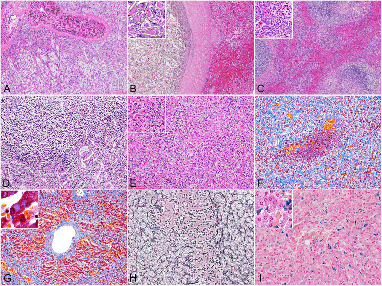
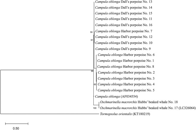
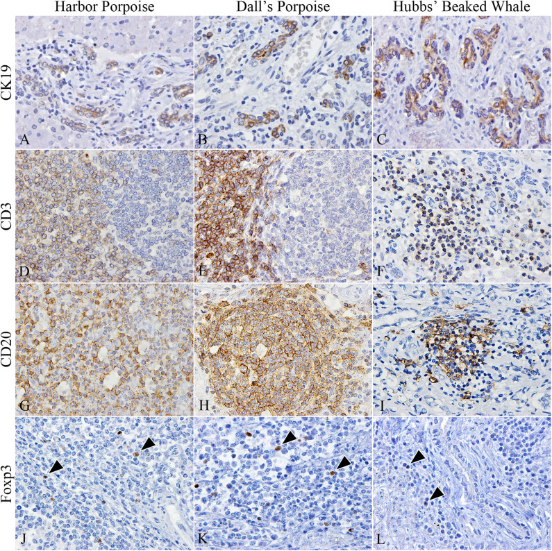
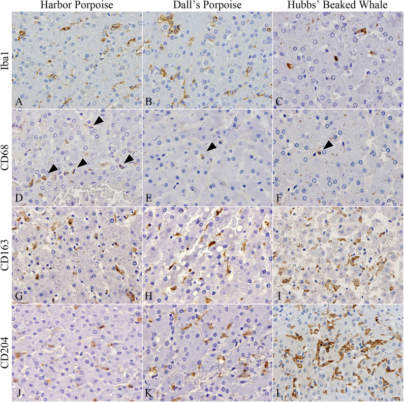
Hepatic trematodiasis is a common condition in a number of free-ranging cetacean species, which occasionally result in severe hepatic and/or pancreatic lesions. However, even the basic pathological information of this disease is unknown for the majority of affected species. The current study describes and compares the histomorphology and immune reaction induced by hepatic trematodes of the family Brachycladiidae in the liver of the harbor porpoise ( = 8), Dall's porpoise ( = 8), and Hubbs' beaked whale ( = 2). Immunohistochemistry for eight antibodies (CK19, CD3, Foxp3, CD20, Iba1, CD68, CD163, and CD204) was conducted to analyze the pathology of these parasitic infections. In all three odontocete species, the changes observed in the trematode-affected biliary epithelium were comparable with marked hyperplasia and goblet cell metaplasia, as well as lymphoplasmacytic and eosinophilic inflammation. Additionally, regions of the Glisson's sheath were diffusely and severely fibrotic in all examined species, regardless of the physical presence of trematodes. Differences among the three species included the presence of characteristic lymphoid follicles formed in the fibrotic bile duct walls of only the two porpoise species. In the Hubbs' beaked whale, the degree of lymphoplasmacytic cholangitis was more severe, and ductular reaction was generally more prominent. In terms of the overall macrophage population among the three species, CD163- and CD204-positive cells (M2 macrophages) outnumbered Iba1- and CD68-positive cells (M1 macrophages), indicating a chronic infection stage in all analyzed individuals. Species-specific differences among the infiltrating macrophages included numbers of CD68-positive cells being significantly more abundant in the harbor porpoises, whereas CD163-positive cells were significantly more numerous in the Dall's porpoises. The numbers of CD204-positive macrophages were higher in the Hubbs' beaked whales compared to those in the porpoises. Trematode species of the harbor and Dall's porpoises were , while they were in the Hubbs' beaked whales. This study concludes that interspecies differences in the tissue reactions to hepatic trematode infections are present among odontocete species and that the immune reaction varies depending on the species. This information aids in furthering our understanding of the pathogenesis of hepatic trematodiasis in cetaceans.
肝吸虫病在许多野生鲸类物种中较为常见,偶尔会导致严重的肝脏和/或胰腺病变。然而,对于大多数受影响的物种来说,这种疾病的基本病理信息尚不清楚。本研究描述并比较了短腺科肝吸虫在港湾鼠海豚(n = 8)、白腰鼠海豚(n = 8)和哈氏喙鲸(n = 2)肝脏中诱导的组织形态学和免疫反应。使用八种抗体(CK19、CD3、Foxp3、CD20、Iba1、CD68、CD163和CD204)进行免疫组织化学分析,以研究这些寄生虫感染的病理学。在所有三种齿鲸物种中,受吸虫影响的胆管上皮细胞变化相似,表现为明显的增生和杯状细胞化生,以及淋巴细胞和嗜酸性粒细胞炎症。此外,在所有检查的物种中,无论是否存在吸虫,肝门管区均出现弥漫性重度纤维化。三种物种之间的差异包括仅在两种鼠海豚的纤维化胆管壁中形成特征性淋巴滤泡。在哈氏喙鲸中,淋巴细胞性胆管炎程度更严重,小胆管反应通常更突出。在三种物种的总体巨噬细胞群体方面,CD163和CD204阳性细胞(M2巨噬细胞)数量超过Iba1和CD68阳性细胞(M1巨噬细胞),表明所有分析个体均处于慢性感染阶段。浸润巨噬细胞的物种特异性差异包括港湾鼠海豚中CD68阳性细胞数量明显更多,而白腰鼠海豚中CD163阳性细胞数量明显更多。哈氏喙鲸中CD204阳性巨噬细胞数量高于鼠海豚。港湾鼠海豚和白腰鼠海豚的吸虫种类为 ,而哈氏喙鲸的为 。本研究得出结论,齿鲸物种对肝吸虫感染的组织反应存在种间差异,免疫反应因物种而异。这些信息有助于加深我们对鲸类肝吸虫病发病机制的理解。