Han Su, Tang Qiaoran, Chen Rui, Li Yihong, Shu Jing, Zhang Xiaoli
Department of Parasitology, Harbin Medical University, Harbin, 150081, China.
Department of Orthopaedic, The fourth Affiliated Hospital of Harbin Medical University, Harbin, China.
BMC Infect Dis. 2017 Aug 1;17(1):531. doi: 10.1186/s12879-017-2630-3.
Hepatic iron overload has been implicated in many liver diseases; however, whether it is involved in clonorchiasis remains unknown. The purpose of this study is to investigate whether Clonorchis sinensis (C. sinensis) infection causes hepatic iron overload, analyze the relationship between the iron overload and associated cell apoptosis, so as to determine the role of excess iron plays in C. sinensis-induced liver injury.
The Perls' Prussian staining and atomic absorption spectrometry methods were used to investigate the iron overload in hepatic sections of wistar rats and patients infected with C. sinensis. The hepatic apoptosis was detected by transferase uridyl nick end labeling (TUNEL) methods. Spearman analysis was used for determining the correlation of the histological hepatic iron index and the apoptotic index.
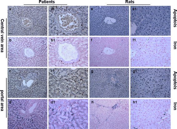
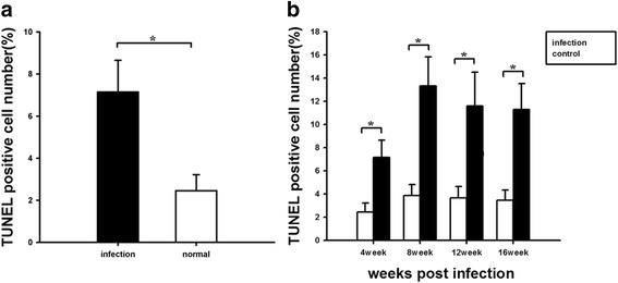
Blue iron particles were deposited mainly in the hepatocytes, Kupffer cells and endothelial cells, around the liver portal and central vein area of both patients and rats. The total iron score was found to be higher in the infected groups than the respective control from 8 weeks. The hepatic iron concentration was also significantly higher in treatment groups than in control rats from 8 weeks. The hepatocyte apoptosis was found to be significantly higher in the portal area of the liver tissue and around the central vein. However, spearman's rank correlation coefficient revealed that there was a mildly negative correlation between the iron index and hepatocyte apoptosis.
This present study confirmed that hepatic iron overload was found during C. sinensis infection. This suggests that iron overload may be associated with hepatocyte apoptosis and involved in liver injury during C. sinensis infection. Further studies are needed to investigate the molecular mechanism involved here.
肝铁过载与多种肝脏疾病有关;然而,其是否参与华支睾吸虫病仍不清楚。本研究的目的是调查华支睾吸虫感染是否会导致肝铁过载,分析铁过载与相关细胞凋亡之间的关系,从而确定过量铁在华支睾吸虫诱导的肝损伤中所起的作用。
采用普鲁士蓝染色法和原子吸收光谱法研究感染华支睾吸虫的Wistar大鼠和患者肝脏切片中的铁过载情况。通过末端脱氧核苷酸转移酶介导的缺口末端标记(TUNEL)法检测肝脏细胞凋亡。采用Spearman分析确定组织学肝铁指数与凋亡指数的相关性。
蓝色铁颗粒主要沉积在患者和大鼠肝脏门静脉和中央静脉区域周围的肝细胞、库普弗细胞和内皮细胞中。从第8周起,发现感染组的总铁评分高于各自的对照组。从第8周起,治疗组的肝脏铁浓度也显著高于对照大鼠。发现肝组织门静脉区域和中央静脉周围的肝细胞凋亡明显增加。然而,Spearman等级相关系数显示铁指数与肝细胞凋亡之间存在轻度负相关。
本研究证实华支睾吸虫感染期间存在肝铁过载。这表明铁过载可能与肝细胞凋亡有关,并参与华支睾吸虫感染期间的肝损伤。需要进一步研究来探讨其中涉及的分子机制。