• 文献检索
  • 文档翻译
  • 深度研究
  • 学术资讯
  • Suppr Zotero 插件Zotero 插件
  • 邀请有礼
  • 套餐&价格
  • 历史记录
应用&插件
Suppr Zotero 插件Zotero 插件浏览器插件Mac 客户端Windows 客户端微信小程序
定价
高级版会员购买积分包购买API积分包
服务
文献检索文档翻译深度研究API 文档MCP 服务
关于我们
关于 Suppr公司介绍联系我们用户协议隐私条款
关注我们

Suppr 超能文献

核心技术专利:CN118964589B侵权必究
粤ICP备2023148730 号-1Suppr @ 2026

文献检索

告别复杂PubMed语法,用中文像聊天一样搜索,搜遍4000万医学文献。AI智能推荐,让科研检索更轻松。

立即免费搜索

文件翻译

保留排版,准确专业,支持PDF/Word/PPT等文件格式,支持 12+语言互译。

免费翻译文档

深度研究

AI帮你快速写综述,25分钟生成高质量综述,智能提取关键信息,辅助科研写作。

立即免费体验

肥厚型心肌病细胞基因表达谱分析揭示病理重构中的新角色。

Gene expression profiling of hypertrophic cardiomyocytes identifies new players in pathological remodelling.

机构信息

Hubrecht Institute, Royal Netherlands Academy of Arts and Sciences and University Medical Center Utrecht, 3584 CT Utrecht, The Netherlands.

Department of Cardiology, University Medical Centre Utrecht, Utrecht, The Netherlands.

出版信息

Cardiovasc Res. 2021 May 25;117(6):1532-1545. doi: 10.1093/cvr/cvaa233.

DOI:10.1093/cvr/cvaa233
PMID:32717063
原文链接:https://pmc.ncbi.nlm.nih.gov/articles/PMC8152696/
Abstract

AIMS

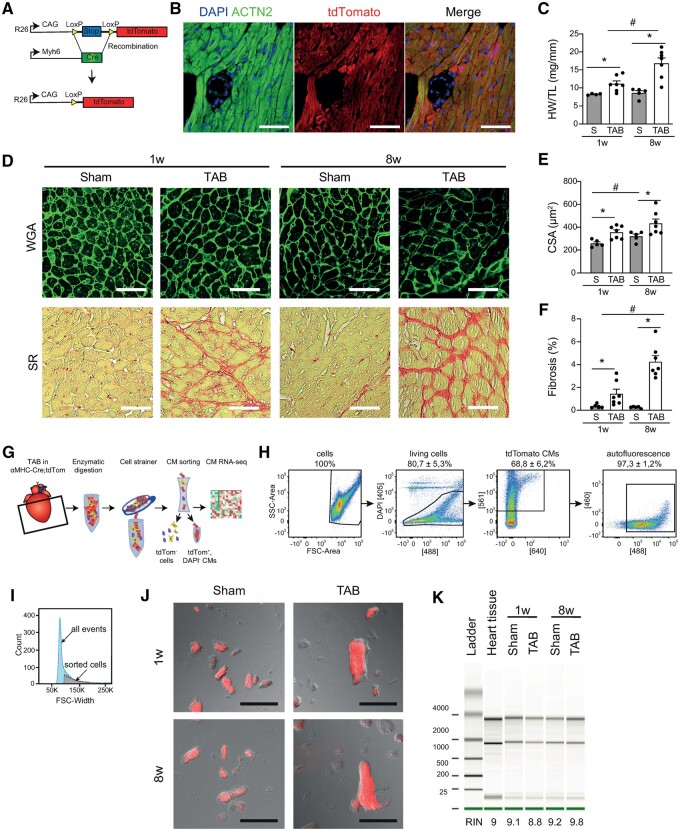
Pathological cardiac remodelling is characterized by cardiomyocyte (CM) hypertrophy and fibroblast activation, which can ultimately lead to maladaptive hypertrophy and heart failure (HF). Genome-wide expression analysis on heart tissue has been instrumental for the identification of molecular mechanisms at play. However, these data were based on signals derived from all cardiac cell types. Here, we aimed for a more detailed view on molecular changes driving maladaptive CM hypertrophy to aid in the development of therapies to reverse pathological remodelling.

METHODS AND RESULTS

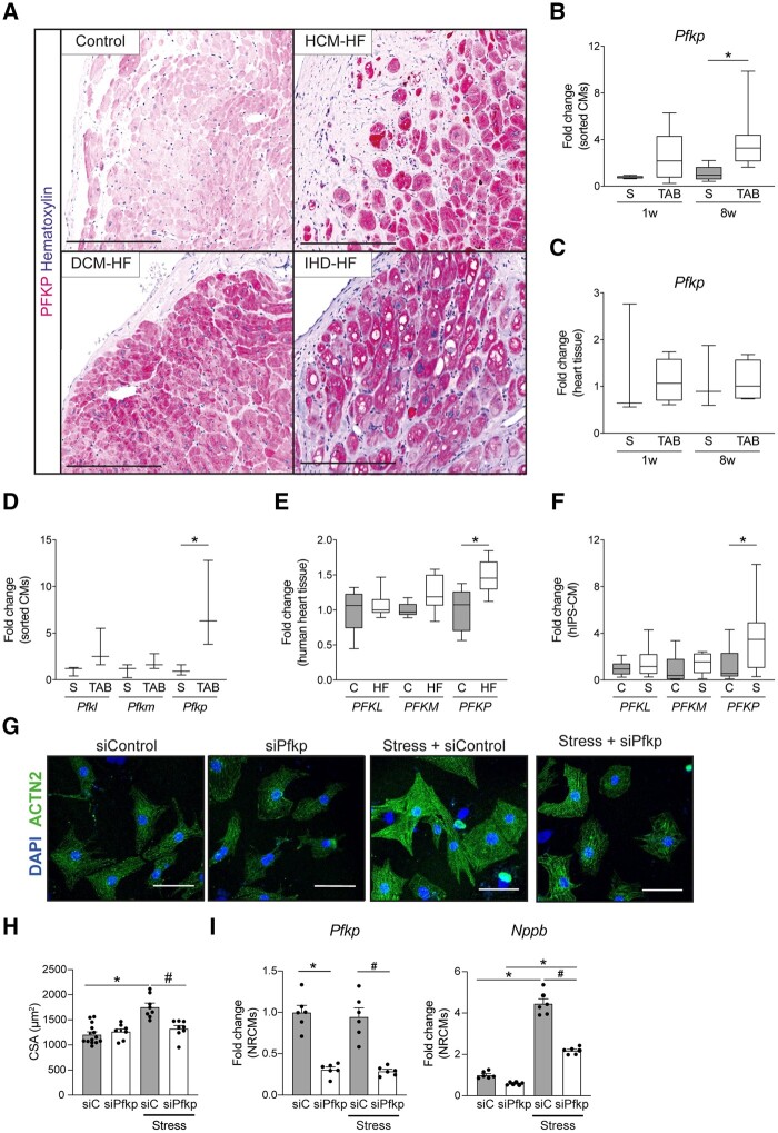
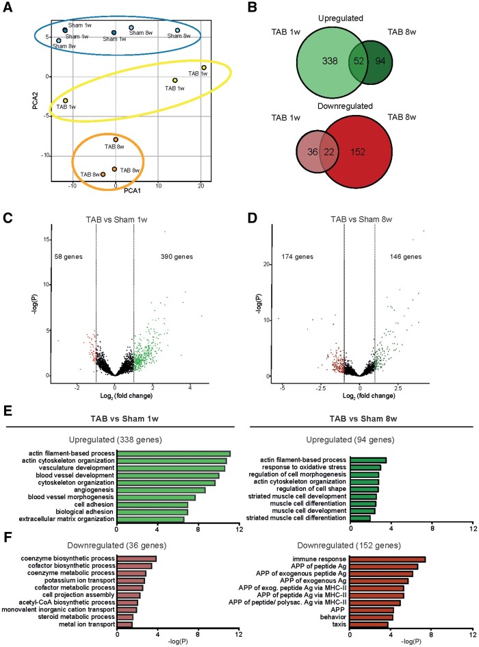
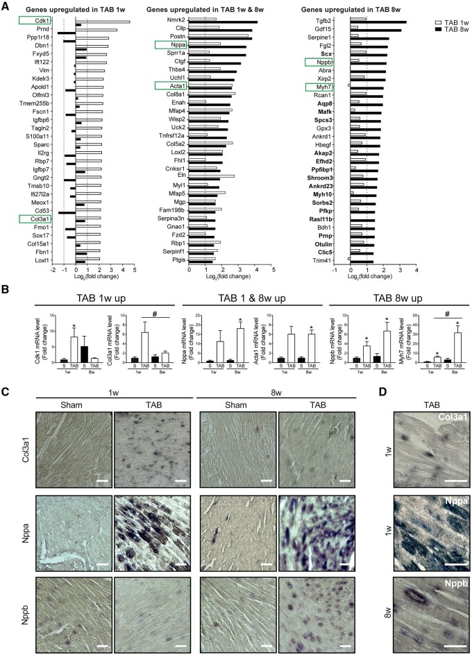
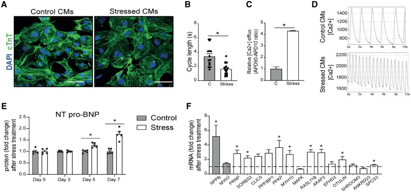
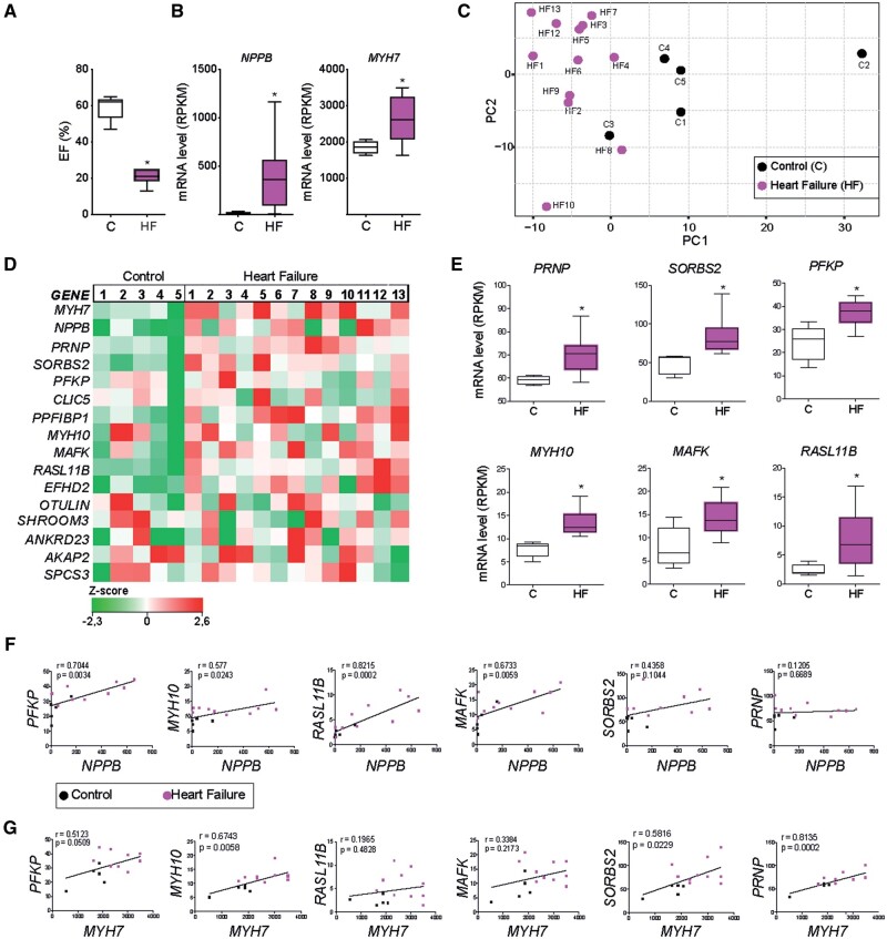
Utilizing CM-specific reporter mice exposed to pressure overload by transverse aortic banding and CM isolation by flow cytometry, we obtained gene expression profiles of hypertrophic CMs in the more immediate phase after stress, and CMs showing pathological hypertrophy. We identified subsets of genes differentially regulated and specific for either stage. Among the genes specifically up-regulated in the CMs during the maladaptive phase we found known stress markers, such as Nppb and Myh7, but additionally identified a set of genes with unknown roles in pathological hypertrophy, including the platelet isoform of phosphofructokinase (PFKP). Norepinephrine-angiotensin II treatment of cultured human CMs induced the secretion of N-terminal-pro-B-type natriuretic peptide (NT-pro-BNP) and recapitulated the up-regulation of these genes, indicating conservation of the up-regulation in failing CMs. Moreover, several genes induced during pathological hypertrophy were also found to be increased in human HF, with their expression positively correlating to the known stress markers NPPB and MYH7. Mechanistically, suppression of Pfkp in primary CMs attenuated stress-induced gene expression and hypertrophy, indicating that Pfkp is an important novel player in pathological remodelling of CMs.

CONCLUSION

Using CM-specific transcriptomic analysis, we identified novel genes induced during pathological hypertrophy that are relevant for human HF, and we show that PFKP is a conserved failure-induced gene that can modulate the CM stress response.

摘要

目的

病理性心脏重构的特征是心肌细胞(CM)肥大和成纤维细胞激活,最终导致适应性不良的肥大和心力衰竭(HF)。对心脏组织进行全基因组表达分析对于鉴定发挥作用的分子机制至关重要。然而,这些数据基于来自所有心肌细胞类型的信号。在这里,我们旨在更详细地观察驱动适应性不良 CM 肥大的分子变化,以帮助开发逆转病理性重构的治疗方法。

方法和结果

利用 CM 特异性报告小鼠通过横主动脉缩窄暴露于压力超负荷,并通过流式细胞术分离 CM,我们获得了应激后更直接阶段的肥大 CM 的基因表达谱,以及表现出病理性肥大的 CM。我们鉴定了差异调节和特定于任一阶段的基因子集。在适应性不良阶段 CM 中特异性上调的基因中,我们发现了已知的应激标志物,如 Nppb 和 Myh7,但另外还发现了一组在病理性肥大中具有未知作用的基因,包括磷酸果糖激酶(PFKP)的血小板同工型。培养的人 CM 中去甲肾上腺素-血管紧张素 II 处理诱导 N 端前 B 型利钠肽(NT-pro-BNP)的分泌,并重新激活这些基因的上调,表明衰竭 CM 中的上调保持不变。此外,在病理性肥大期间诱导的几个基因也在人类 HF 中增加,其表达与已知的应激标志物 NPPB 和 MYH7 呈正相关。在机制上,在原代 CM 中抑制 Pfkp 可减弱应激诱导的基因表达和肥大,表明 Pfkp 是 CM 病理性重构的一个重要新的调节因子。

结论

使用 CM 特异性转录组分析,我们鉴定了在病理性肥大期间诱导的与人类 HF 相关的新基因,并且我们表明 PFKP 是一个保守的衰竭诱导基因,可以调节 CM 的应激反应。

https://cdn.ncbi.nlm.nih.gov/pmc/blobs/34b6/8152696/b4561c70c798/cvaa233f6.jpg
https://cdn.ncbi.nlm.nih.gov/pmc/blobs/34b6/8152696/10d116bc21ec/cvaa233f1.jpg
https://cdn.ncbi.nlm.nih.gov/pmc/blobs/34b6/8152696/468a3b481f71/cvaa233f2.jpg
https://cdn.ncbi.nlm.nih.gov/pmc/blobs/34b6/8152696/65f096d62456/cvaa233f3.jpg
https://cdn.ncbi.nlm.nih.gov/pmc/blobs/34b6/8152696/2105d2e01632/cvaa233f4.jpg
https://cdn.ncbi.nlm.nih.gov/pmc/blobs/34b6/8152696/509e6d95a1e0/cvaa233f5.jpg
https://cdn.ncbi.nlm.nih.gov/pmc/blobs/34b6/8152696/b4561c70c798/cvaa233f6.jpg

相似文献

1
Gene expression profiling of hypertrophic cardiomyocytes identifies new players in pathological remodelling.肥厚型心肌病细胞基因表达谱分析揭示病理重构中的新角色。
Cardiovasc Res. 2021 May 25;117(6):1532-1545. doi: 10.1093/cvr/cvaa233.
2
Neuron-derived orphan receptor-1 modulates cardiac gene expression and exacerbates angiotensin II-induced cardiac hypertrophy.神经元衍生孤儿受体 1 调节心脏基因表达并加剧血管紧张素 II 诱导的心脏肥大。
Clin Sci (Lond). 2020 Feb 14;134(3):359-377. doi: 10.1042/CS20191014.
3
Angiotensin II type-1 receptor activation in the adult heart causes blood pressure-independent hypertrophy and cardiac dysfunction.成年心脏中血管紧张素II 1型受体的激活会导致与血压无关的心肌肥大和心脏功能障碍。
Cardiovasc Res. 2009 Feb 15;81(3):592-600. doi: 10.1093/cvr/cvn230. Epub 2008 Aug 14.
4
A state of reversible compensated ventricular dysfunction precedes pathological remodelling in response to cardiomyocyte-specific activity of angiotensin II type-1 receptor in mice.在小鼠中,血管紧张素II 1型受体的心肌细胞特异性活性引起的病理性重塑之前,存在可逆性代偿性心室功能障碍状态。
Dis Model Mech. 2015 Aug 1;8(8):783-94. doi: 10.1242/dmm.019174. Epub 2015 Jun 18.
5
Qiliqiangxin Attenuates Phenylephrine-Induced Cardiac Hypertrophy through Downregulation of MiR-199a-5p.芪苈强心通过下调miR-199a-5p减轻去甲肾上腺素诱导的心肌肥厚。
Cell Physiol Biochem. 2016;38(5):1743-51. doi: 10.1159/000443113. Epub 2016 May 9.
6
Adiponectin mediates cardioprotection in oxidative stress-induced cardiac myocyte remodeling.脂联素介导氧化应激诱导的心肌细胞重构中的心脏保护作用。
Am J Physiol Heart Circ Physiol. 2011 Sep;301(3):H984-93. doi: 10.1152/ajpheart.00428.2011. Epub 2011 Jun 10.
7
NF-kappaB activation is required for adaptive cardiac hypertrophy.NF-κB 的激活对于适应性心肌肥厚是必需的。
Cardiovasc Res. 2009 Dec 1;84(3):416-24. doi: 10.1093/cvr/cvp237. Epub 2009 Jul 20.
8
Attenuation of Cardiomyocyte Hypertrophy via Depletion Myh7 using CASAAV.利用腺相关病毒相关的CRISPR/Cas9系统敲低Myh7基因以减轻心肌细胞肥大
Cardiovasc Toxicol. 2021 Mar;21(3):255-264. doi: 10.1007/s12012-020-09617-y. Epub 2020 Oct 23.
9
Mixed lineage kinase-3 prevents cardiac dysfunction and structural remodeling with pressure overload.混合谱系激酶-3 可预防心脏压力超负荷导致的心脏功能障碍和结构重塑。
Am J Physiol Heart Circ Physiol. 2019 Jan 1;316(1):H145-H159. doi: 10.1152/ajpheart.00029.2018. Epub 2018 Oct 26.
10
MicroRNA-27b-3p down-regulates FGF1 and aggravates pathological cardiac remodelling.miRNA-27b-3p 下调 FGF1 并加重病理性心脏重构。
Cardiovasc Res. 2022 Jul 20;118(9):2139-2151. doi: 10.1093/cvr/cvab248.

引用本文的文献

1
ArfGAP with Dual Pleckstrin Homology Domains 2 Promotes Hypertrophy of Cultured Neonatal Cardiomyocytes.具有双pleckstrin同源结构域2的ArfGAP促进培养的新生心肌细胞肥大。
Int J Mol Sci. 2025 Aug 6;26(15):7588. doi: 10.3390/ijms26157588.
2
Gen-miR-5 derived from inhibits PFKP to prevent fibroblast activation and alleviate myocardial fibrosis.源自 的基因微小RNA-5抑制磷酸果糖激酶P以防止成纤维细胞活化并减轻心肌纤维化。
Front Pharmacol. 2025 May 2;16:1578877. doi: 10.3389/fphar.2025.1578877. eCollection 2025.
3
Regulatory roles of PIWI-interacting RNAs in cardiovascular disease.

本文引用的文献

1
Mid-regional pro-atrial natriuretic peptide for the early detection of non-acute heart failure.中段心房利钠肽原用于早期检测非急性心力衰竭。
Eur J Heart Fail. 2019 Oct;21(10):1219-1227. doi: 10.1002/ejhf.1495. Epub 2019 Jun 17.
2
Cardiomyocytes facing fibrotic conditions re-express extracellular matrix transcripts.心肌细胞在面对纤维化条件时会重新表达细胞外基质的转录本。
Acta Biomater. 2019 Apr 15;89:180-192. doi: 10.1016/j.actbio.2019.03.017. Epub 2019 Mar 9.
3
Single-Cell Transcriptome Analysis Maps the Developmental Track of the Human Heart.
PIWI相互作用RNA在心血管疾病中的调控作用。
Am J Physiol Heart Circ Physiol. 2025 Apr 1;328(4):H991-H1004. doi: 10.1152/ajpheart.00833.2024. Epub 2025 Mar 6.
4
The role of mechanosignaling in the control of myocardial mass.机械信号传导在心肌质量控制中的作用。
Am J Physiol Heart Circ Physiol. 2025 Mar 1;328(3):H622-H638. doi: 10.1152/ajpheart.00277.2024. Epub 2024 Dec 31.
5
Potential diagnostic value of circulating miRNAs in HFrEF and bioinformatics analysis.循环微小RNA在射血分数降低的心力衰竭中的潜在诊断价值及生物信息学分析
Heliyon. 2024 Sep 20;10(19):e37929. doi: 10.1016/j.heliyon.2024.e37929. eCollection 2024 Oct 15.
6
Gene expression profiles, potential targets and treatments of cardiac remodeling.心脏重塑的基因表达谱、潜在靶点及治疗方法
Mol Cell Biochem. 2025 Mar;480(3):1555-1567. doi: 10.1007/s11010-024-05126-6. Epub 2024 Oct 5.
7
PRDM16 determines specification of ventricular cardiomyocytes by suppressing alternative cell fates.PRDM16 通过抑制心脏细胞的其他命运决定来确定心室肌细胞的特征。
Life Sci Alliance. 2024 Sep 20;7(12). doi: 10.26508/lsa.202402719. Print 2024 Dec.
8
Opa1 processing is dispensable in mouse development but is protective in mitochondrial cardiomyopathy.OPA1 加工在小鼠发育中是可有可无的,但在心肌线粒体病中具有保护作用。
Sci Adv. 2024 Aug 2;10(31):eadp0443. doi: 10.1126/sciadv.adp0443.
9
Cardiomyocyte intercellular signalling increases oxidative stress and reprograms the global- and phospho-proteome of cardiac fibroblasts.心肌细胞间信号传导增加氧化应激并对心脏成纤维细胞的整体蛋白质组和磷酸化蛋白质组进行重编程。
J Extracell Biol. 2023 Nov 30;2(12):e125. doi: 10.1002/jex2.125. eCollection 2023 Dec.
10
Combined Pharmacological Modulation of Translational and Transcriptional Activity Signaling Pathways as a Promising Therapeutic Approach in Children with Myocardial Changes.联合药理学调节翻译和转录活性信号通路作为治疗心肌改变儿童的一种有前景的治疗方法。
Biomolecules. 2024 Apr 13;14(4):477. doi: 10.3390/biom14040477.
单细胞转录组分析描绘了人类心脏的发育轨迹。
Cell Rep. 2019 Feb 12;26(7):1934-1950.e5. doi: 10.1016/j.celrep.2019.01.079.
4
Cardiomyocyte gene programs encoding morphological and functional signatures in cardiac hypertrophy and failure.心肌细胞基因程序在心肌肥厚和衰竭中编码形态和功能特征。
Nat Commun. 2018 Oct 30;9(1):4435. doi: 10.1038/s41467-018-06639-7.
5
Study on the mechanism behind lncRNA MEG3 affecting clear cell renal cell carcinoma by regulating miR-7/RASL11B signaling.探讨长链非编码 RNA MEG3 通过调控 miR-7/RASL11B 信号通路影响肾透明细胞癌的作用机制。
J Cell Physiol. 2018 Dec;233(12):9503-9515. doi: 10.1002/jcp.26849. Epub 2018 Jul 3.
6
Metabolic remodelling in heart failure.心力衰竭中的代谢重编程。
Nat Rev Cardiol. 2018 Aug;15(8):457-470. doi: 10.1038/s41569-018-0044-6.
7
Single-Cell Sequencing of the Healthy and Diseased Heart Reveals Cytoskeleton-Associated Protein 4 as a New Modulator of Fibroblasts Activation.单细胞测序技术揭示健康和病变心脏中的细胞骨架相关蛋白 4 是成纤维细胞激活的新型调节剂。
Circulation. 2018 Jul 10;138(2):166-180. doi: 10.1161/CIRCULATIONAHA.117.030742. Epub 2018 Jan 31.
8
Gene expression analysis to identify mechanisms underlying heart failure susceptibility in mice and humans.基因表达分析鉴定小鼠和人类心力衰竭易感性的潜在机制。
Basic Res Cardiol. 2017 Dec 29;113(1):8. doi: 10.1007/s00395-017-0666-6. Print 2018 Jan 8.
9
Stabilization of phosphofructokinase 1 platelet isoform by AKT promotes tumorigenesis.AKT 稳定磷酸果糖激酶 1 血小板同工型促进肿瘤发生。
Nat Commun. 2017 Oct 16;8(1):949. doi: 10.1038/s41467-017-00906-9.
10
Cellular Communications in the Heart.心脏中的细胞通讯
Card Fail Rev. 2015 Oct;1(2):64-68. doi: 10.15420/cfr.2015.1.2.64.