Li Jiali, Xun Yang, Li Cong, Han Yunfeng, Shen Yaqi, Hu Xuemei, Hu Daoyu, Liu Zheng, Wang Shaogang, Li Zhen
Department of Radiology, Tongji Hospital, Tongji Medical College, Huazhong University of Science and Technology, Wuhan, China.
Department of Urology, Tongji Hospital, Tongji Medical College, Huazhong University of Science and Technology, Wuhan, China.
Front Med (Lausanne). 2020 Jul 3;7:309. doi: 10.3389/fmed.2020.00309. eCollection 2020.
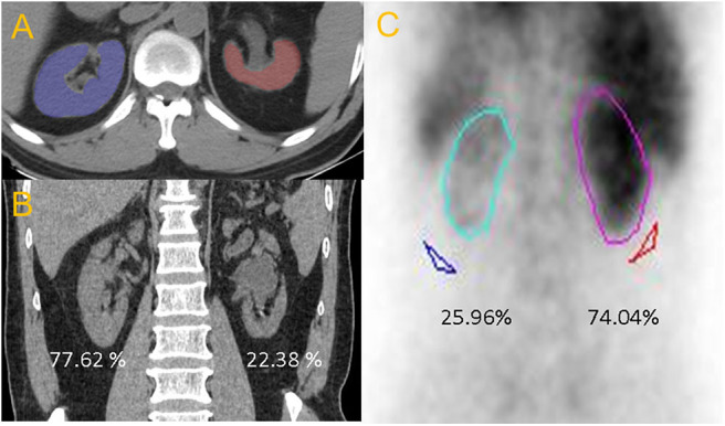
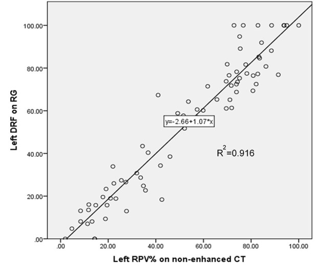
The aim of this study was to determine whether unenhanced computed tomography (CT) imaging can estimate differential renal function (DRF) in patients with chronic unilateral obstructive upper urinary tract stones. This was a single-center retrospective study of 76 patients. All the patients underwent unenhanced CT and nuclear renography (RG) at an interval of 4 to 6 weeks due to chronic unilateral obstructive urinary stones. Renal CT measurements (RCMs), including residual parenchymal volume (RPV) and volumetric CT texture analysis parameters, were obtained through a semiautomatic method. Percent RCMs were calculated and compared with renal function determined by RG. The strongest Pearson coefficient between percent RCM and DRF was reflected by RPV ( = 0.957, < 0.001). Combinations of RPV and other parameters did not significantly improve the correlation compared with RPV alone ( = 0.957 vs. = 0.957, 0.957, 0.887, 0.815, and 0.956 for combination with Hounsfield unit, parenchymal voxel, skewness, kurtosis, and entropy, respectively; all < 0.001). Percent RPV was subsequently introduced into linear regression, and the equation y = -2.66 + 1.07 × ( < 0.001) was derived to calculate predicted DRF. No statistically difference was found between predicted DRF using the equation and observed DRF according to RG ( = 0.959). Unenhanced CT imaging can estimate DRF in patients with chronic unilateral obstructive upper urinary tract stones, and RG might not be necessary as a conventional method in clinical.
本研究的目的是确定非增强计算机断层扫描(CT)成像能否评估慢性单侧梗阻性上尿路结石患者的分肾功能(DRF)。这是一项对76例患者进行的单中心回顾性研究。由于慢性单侧梗阻性尿路结石,所有患者均在4至6周的间隔内接受了非增强CT和核肾图(RG)检查。通过半自动方法获得肾脏CT测量值(RCMs),包括残余实质体积(RPV)和体积CT纹理分析参数。计算RCMs百分比,并与通过RG测定的肾功能进行比较。RPV反映了RCM百分比与DRF之间最强的Pearson系数(= 0.957,< 0.001)。与单独的RPV相比,RPV与其他参数的组合并未显著改善相关性(与Hounsfield单位、实质体素、偏度、峰度和熵组合时分别为= 0.957 vs. = 0.957、0.957、0.887、0.815和0.956;均< 0.001)。随后将RPV百分比引入线性回归,得出方程y = -2.66 + 1.07 ×(< 0.001)以计算预测的DRF。使用该方程预测的DRF与根据RG观察到的DRF之间未发现统计学差异(= 0.959)。非增强CT成像可以评估慢性单侧梗阻性上尿路结石患者的DRF,并且在临床中可能无需将RG作为常规方法。