Hasegawa Daigaku, Hasegawa Kana, Kaneko Hiroshi, Yoshida Shinichiro, Mitarai Hiromi, Arima Mai, Tomokiyo Atsushi, Hamano Sayuri, Sugii Hideki, Wada Naohisa, Kiyoshima Tamotsu, Maeda Hidefumi
Department of Endodontology, Kyushu University Hospital, Kyushu University, 3-1-1 Maidashi, Higashi-ku, Fukuoka 812-8582, Japan.
Department of Oral Pathology, Faculty of Dental Science, Kyushu University, 3-1-1 Maidashi, Higashi-ku, Fukuoka 812-8582, Japan.
Stem Cells Int. 2020 Jul 8;2020:9672673. doi: 10.1155/2020/9672673. eCollection 2020.
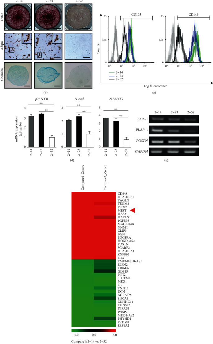
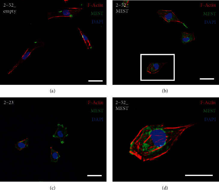
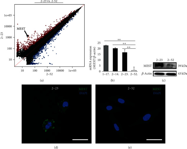
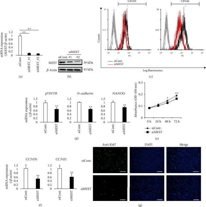
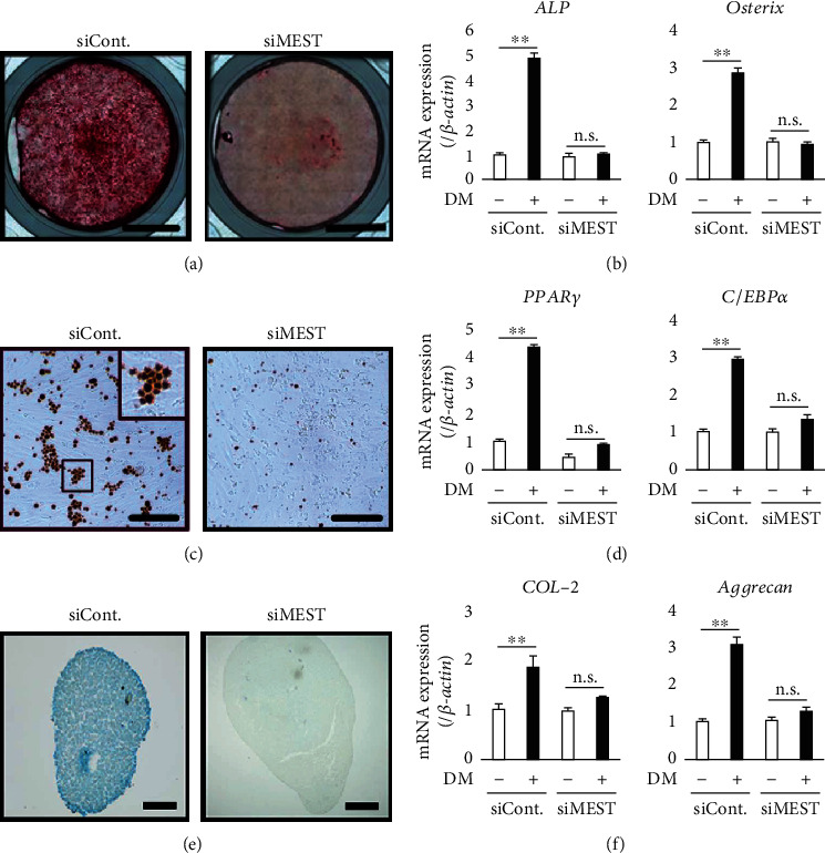
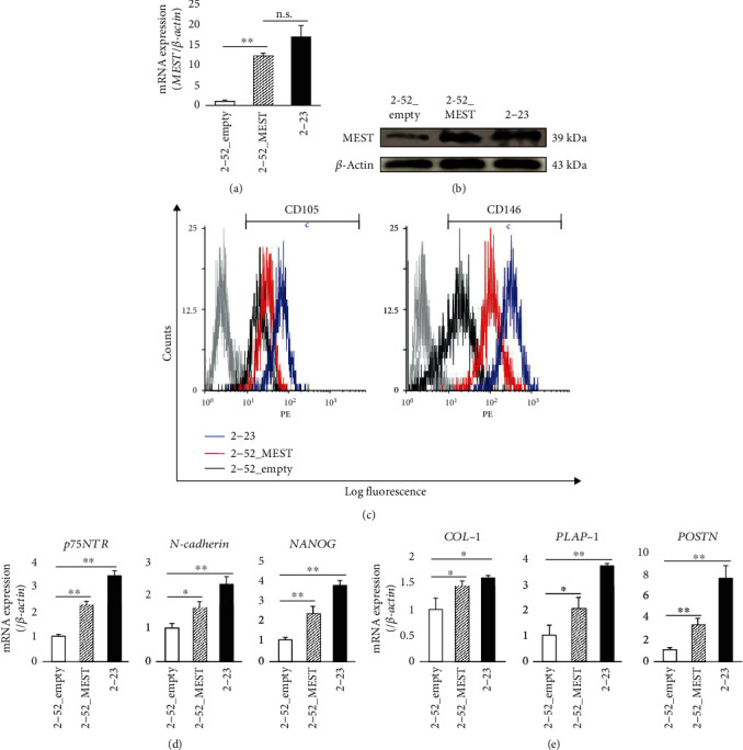
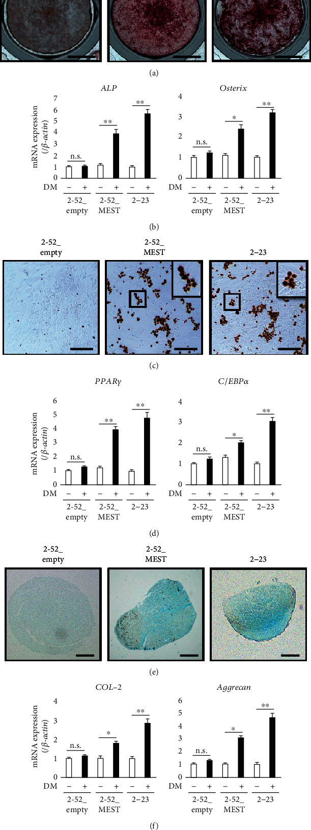
Periodontal ligament (PDL) stem cells (PDLSCs) have been reported as a useful cell source for periodontal tissue regeneration. However, one of the issues is the difficulty of obtaining a sufficient number of PDLSCs for clinical application because very few PDLSCs can be isolated from PDL tissue of donors. Therefore, we aimed to identify a specific factor that converts human PDL cells into stem-like cells. In this study, microarray analysis comparing the gene profiles of human PDLSC lines (2-14 and 2-23) with those of a cell line with a low differentiation potential (2-52) identified the imprinted gene mesoderm-specific transcript (MEST). MEST was expressed in the cytoplasm of 2-23 cells. Knockdown of MEST by siRNA in 2-23 cells inhibited the expression of stem cell markers, such as CD105, CD146, p75NTR, N-cadherin, and NANOG; the proliferative potential; and multidifferentiation capacity for osteoblasts, adipocytes, and chondrocytes. On the other hand, overexpression of MEST in 2-52 cells enhanced the expression of stem cell markers and PDL-related markers and the multidifferentiation capacity. In addition, MEST-overexpressing 2-52 cells exhibited a change in morphology from a spindle shape to a stem cell-like round shape that was similar to 2-14 and 2-23 cell morphologies. These results suggest that MEST plays a critical role in the maintenance of stemness in PDLSCs and converts PDL cells into PDLSC-like cells. Therefore, this study indicates that MEST may be a therapeutic factor for periodontal tissue regeneration by inducing PDLSCs.
牙周膜(PDL)干细胞(PDLSCs)已被报道为牙周组织再生的一种有用细胞来源。然而,问题之一是难以获得足够数量的PDLSCs用于临床应用,因为从供体的PDL组织中只能分离出很少的PDLSCs。因此,我们旨在确定一种将人PDL细胞转化为干细胞样细胞的特定因子。在本研究中,通过微阵列分析比较人PDLSC系(2-14和2-23)与低分化潜能细胞系(2-52)的基因谱,鉴定出印记基因中胚层特异性转录本(MEST)。MEST在2-23细胞的细胞质中表达。在2-23细胞中用siRNA敲低MEST可抑制干细胞标志物如CD105、CD146、p75NTR、N-钙黏蛋白和NANOG的表达、增殖潜能以及向成骨细胞、脂肪细胞和软骨细胞的多分化能力。另一方面,在2-52细胞中过表达MEST可增强干细胞标志物和PDL相关标志物的表达以及多分化能力。此外,过表达MEST的2-52细胞形态从纺锤形变为类似于2-14和2-23细胞形态的干细胞样圆形。这些结果表明MEST在维持PDLSCs的干性中起关键作用,并将PDL细胞转化为PDLSC样细胞。因此,本研究表明MEST可能是通过诱导PDLSCs实现牙周组织再生的治疗因子。