• 文献检索
  • 文档翻译
  • 深度研究
  • 学术资讯
  • Suppr Zotero 插件Zotero 插件
  • 邀请有礼
  • 套餐&价格
  • 历史记录
应用&插件
Suppr Zotero 插件Zotero 插件浏览器插件Mac 客户端Windows 客户端微信小程序
定价
高级版会员购买积分包购买API积分包
服务
文献检索文档翻译深度研究API 文档MCP 服务
关于我们
关于 Suppr公司介绍联系我们用户协议隐私条款
关注我们

Suppr 超能文献

核心技术专利:CN118964589B侵权必究
粤ICP备2023148730 号-1Suppr @ 2026

文献检索

告别复杂PubMed语法,用中文像聊天一样搜索,搜遍4000万医学文献。AI智能推荐,让科研检索更轻松。

立即免费搜索

文件翻译

保留排版,准确专业,支持PDF/Word/PPT等文件格式,支持 12+语言互译。

免费翻译文档

深度研究

AI帮你快速写综述,25分钟生成高质量综述,智能提取关键信息,辅助科研写作。

立即免费体验

使用扩散相关成像技术直接且特异性评估轴突损伤和脊髓微环境。

Direct and specific assessment of axonal injury and spinal cord microenvironments using diffusion correlation imaging.

机构信息

The Eunice Kennedy Shriver National Institute of Child Health and Human Development, Bethesda, MD 20817, USA; The Center for Neuroscience and Regenerative Medicine, Uniformed Service University of the Health Sciences, Bethesda, MD 20814, USA.

The Department of Biomedical Engineering, The University of Arizona, Tucson, Arizona 85721, USA.

出版信息

Neuroimage. 2020 Nov 1;221:117195. doi: 10.1016/j.neuroimage.2020.117195. Epub 2020 Jul 26.

DOI:10.1016/j.neuroimage.2020.117195
PMID:32726643
原文链接:https://pmc.ncbi.nlm.nih.gov/articles/PMC7805019/
Abstract

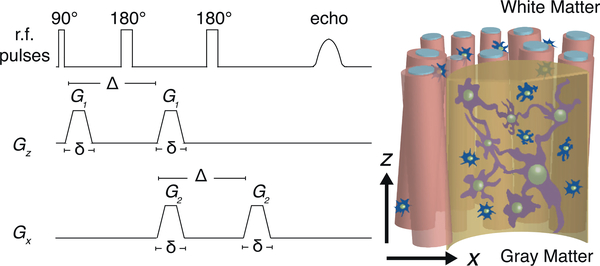
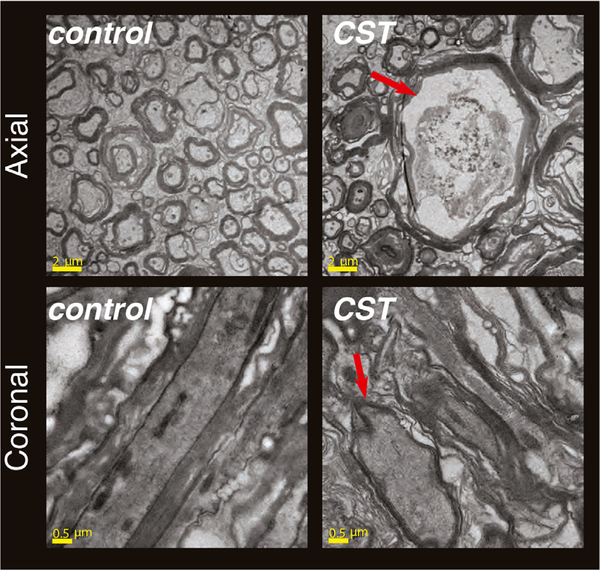
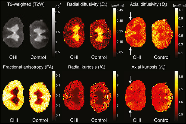
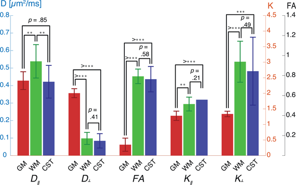
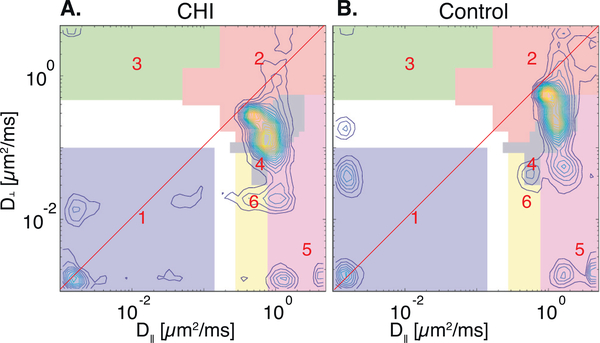
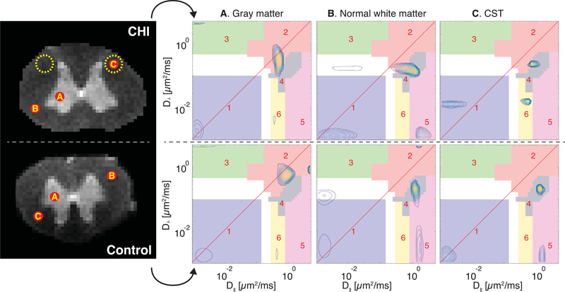
We describe a practical two-dimensional (2D) diffusion MRI framework to deliver specificity and improve sensitivity to axonal injury in the spinal cord. This approach provides intravoxel distributions of correlations of water mobilities in orthogonal directions, revealing sub-voxel diffusion components. Here we use it to investigate water diffusivities along axial and radial orientations within spinal cord specimens with confirmed, tract-specific axonal injury. First, we show using transmission electron microscopy and immunohistochemistry that tract-specific axonal beading occurs following Wallerian degeneration in the cortico-spinal tract as direct sequelae to closed head injury. We demonstrate that although some voxel-averaged diffusion tensor imaging (DTI) metrics are sensitive to this axonal injury, they are non-specific, i.e., they do not reveal an underlying biophysical mechanism of injury. Then we employ 2D diffusion correlation imaging (DCI) to improve discrimination of different water microenvironments by measuring and mapping the joint water mobility distributions perpendicular and parallel to the spinal cord axis. We determine six distinct diffusion spectral components that differ according to their microscopic anisotropy and mobility. We show that at the injury site a highly anisotropic diffusion component completely disappears and instead becomes more isotropic. Based on these findings, an injury-specific MR image of the spinal cord was generated, and a radiological-pathological correlation with histological silver staining % area was performed. The resulting strong and significant correlation (r=0.70,p < 0.0001) indicates the high specificity with which DCI detects injury-induced tissue alterations. We predict that the ability to selectively image microstructural changes following axonal injury in the spinal cord can be useful in clinical and research applications by enabling specific detection and increased sensitivity to injury-induced microstructural alterations. These results also encourage us to translate DCI to higher spatial dimensions to enable assessment of traumatic axonal injury, and possibly other diseases and disorders in the brain.

摘要

我们描述了一种实用的二维(2D)扩散 MRI 框架,以提供特异性并提高脊髓轴突损伤的敏感性。该方法提供了各向异性水迁移率的体素内分布,揭示了亚体素扩散分量。在这里,我们使用它来研究具有明确的、束特异性轴突损伤的脊髓标本中沿轴向和径向方向的水扩散率。首先,我们通过透射电子显微镜和免疫组织化学显示,皮质脊髓束中的束特异性轴突珠状形成发生在沃勒变性之后,这是闭合性颅脑损伤的直接后果。我们证明,尽管一些体素平均扩散张量成像(DTI)指标对这种轴突损伤敏感,但它们是非特异性的,即它们不能揭示损伤的潜在生物物理机制。然后,我们采用 2D 扩散相关成像(DCI)通过测量和绘制与脊髓轴垂直和平行的水迁移率分布来改善对不同水微环境的区分。我们确定了六个不同的扩散光谱分量,它们根据其微观各向异性和迁移率而有所不同。我们表明,在损伤部位,高度各向异性的扩散分量完全消失,取而代之的是更各向同性的扩散分量。基于这些发现,生成了脊髓的损伤特异性 MR 图像,并与组织银染色%面积进行了放射病理学相关性分析。结果表明,相关性很强且具有统计学意义(r=0.70,p<0.0001),这表明 DCI 检测损伤诱导的组织改变具有很高的特异性。我们预测,选择性成像脊髓轴突损伤后微观结构变化的能力可以通过特异性检测和增加对损伤诱导的微观结构改变的敏感性,在临床和研究应用中非常有用。这些结果还鼓励我们将 DCI 转化为更高的空间维度,以评估创伤性轴突损伤,以及可能的大脑中的其他疾病和障碍。

https://cdn.ncbi.nlm.nih.gov/pmc/blobs/8408/7805019/b2062bf29fb3/nihms-1657534-f0009.jpg
https://cdn.ncbi.nlm.nih.gov/pmc/blobs/8408/7805019/15a64a2d915c/nihms-1657534-f0001.jpg
https://cdn.ncbi.nlm.nih.gov/pmc/blobs/8408/7805019/7f6e1f5026b9/nihms-1657534-f0002.jpg
https://cdn.ncbi.nlm.nih.gov/pmc/blobs/8408/7805019/ef6de4a17176/nihms-1657534-f0003.jpg
https://cdn.ncbi.nlm.nih.gov/pmc/blobs/8408/7805019/d50c3f629ae0/nihms-1657534-f0004.jpg
https://cdn.ncbi.nlm.nih.gov/pmc/blobs/8408/7805019/676387701958/nihms-1657534-f0005.jpg
https://cdn.ncbi.nlm.nih.gov/pmc/blobs/8408/7805019/0faee45ae8bf/nihms-1657534-f0006.jpg
https://cdn.ncbi.nlm.nih.gov/pmc/blobs/8408/7805019/05cd1d1b4eba/nihms-1657534-f0007.jpg
https://cdn.ncbi.nlm.nih.gov/pmc/blobs/8408/7805019/5abae498a58b/nihms-1657534-f0008.jpg
https://cdn.ncbi.nlm.nih.gov/pmc/blobs/8408/7805019/b2062bf29fb3/nihms-1657534-f0009.jpg

相似文献

1
Direct and specific assessment of axonal injury and spinal cord microenvironments using diffusion correlation imaging.使用扩散相关成像技术直接且特异性评估轴突损伤和脊髓微环境。
Neuroimage. 2020 Nov 1;221:117195. doi: 10.1016/j.neuroimage.2020.117195. Epub 2020 Jul 26.
2
Wallerian Degeneration Assessed by Multi-Modal Magnetic Resonance Imaging of Cervical Spinal Cord Is Associated With Neurological Impairment After Spinal Cord Injury.通过颈椎脊髓多模态磁共振成像评估的沃勒变性与脊髓损伤后的神经功能障碍相关。
J Neurotrauma. 2024 May;41(9-10):1240-1252. doi: 10.1089/neu.2023.0305. Epub 2024 Apr 15.
3
Wallerian degeneration in cervical spinal cord tracts is commonly seen in routine T2-weighted MRI after traumatic spinal cord injury and is associated with impairment in a retrospective study.颈髓束的沃勒氏变性在创伤性脊髓损伤后的常规 T2 加权 MRI 中很常见,并且与回顾性研究中的功能障碍有关。
Eur Radiol. 2021 May;31(5):2923-2932. doi: 10.1007/s00330-020-07388-2. Epub 2020 Oct 30.
4
Diffusion Tensor Imaging扩散张量成像
5
Diffusion tensor imaging detects early Wallerian degeneration of the pyramidal tract after ischemic stroke.弥散张量成像可检测缺血性卒中后锥体束的早期华勒氏变性。
Neuroimage. 2004 Aug;22(4):1767-74. doi: 10.1016/j.neuroimage.2004.03.041.
6
Characterizing white matter damage in rat spinal cord with quantitative MRI and histology.利用定量磁共振成像和组织学方法表征大鼠脊髓中的白质损伤。
J Neurotrauma. 2008 Jun;25(6):653-76. doi: 10.1089/neu.2007.0462.
7
Wallerian Degeneration Beyond the Corticospinal Tracts: Conventional and Advanced MRI Findings.皮质脊髓束以外的华勒氏变性:传统及先进的磁共振成像表现
J Neuroimaging. 2017 May;27(3):272-280. doi: 10.1111/jon.12404. Epub 2016 Nov 7.
8
Axonal integrity predicts cortical reorganisation following cervical injury.轴突完整性预测颈椎损伤后的皮质重组。
J Neurol Neurosurg Psychiatry. 2012 Jun;83(6):629-37. doi: 10.1136/jnnp-2011-301875. Epub 2012 Apr 6.
9
Diffusion tensor imaging as a biomarker for assessing neuronal stem cell treatments affecting areas distal to the site of spinal cord injury.扩散张量成像作为一种生物标志物,用于评估影响脊髓损伤部位远端区域的神经元干细胞治疗效果。
J Neurosurg Spine. 2017 Feb;26(2):243-251. doi: 10.3171/2016.5.SPINE151319. Epub 2016 Sep 30.
10
Retrograde Wallerian degeneration of cranial corticospinal tracts in cervical spinal cord injury patients using diffusion tensor imaging.利用扩散张量成像技术对颈脊髓损伤患者颅侧皮质脊髓束的逆行性华勒氏变性进行研究。
J Neurosci Res. 2008 Aug 1;86(10):2271-80. doi: 10.1002/jnr.21664.

引用本文的文献

1
Low-field, high-gradient NMR shows diffusion contrast consistent with localization or motional averaging of water near surfaces.低场、高梯度核磁共振显示出与表面附近水的定位或运动平均一致的扩散对比。
Magn Reson Lett. 2023 Apr 8;3(2):90-107. doi: 10.1016/j.mrl.2023.03.009. eCollection 2023 May.
2
Identification of diffusion, kurtosis, and propagator MRI markers of Alzheimer's disease pathology in post-mortem human tissue.在死后人体组织中鉴定阿尔茨海默病病理学的扩散、峰度和传播子磁共振成像标志物。
Imaging Neurosci (Camb). 2024 May 8;2. doi: 10.1162/imag_a_00164. eCollection 2024.
3
Variability of multidimensional diffusion-relaxation MRI estimates in the human brain.

本文引用的文献

1
SANDI: A compartment-based model for non-invasive apparent soma and neurite imaging by diffusion MRI.桑迪:一种基于体素的扩散 MRI 无创表观体素和神经丝成像模型。
Neuroimage. 2020 Jul 15;215:116835. doi: 10.1016/j.neuroimage.2020.116835. Epub 2020 Apr 11.
2
Retaining information from multidimensional correlation MRI using a spectral regions of interest generator.利用谱区兴趣生成器从多维相关 MRI 中保留信息。
Sci Rep. 2020 Feb 24;10(1):3246. doi: 10.1038/s41598-020-60092-5.
3
The dot-compartment revealed? Diffusion MRI with ultra-strong gradients and spherical tensor encoding in the living human brain.
人脑多维扩散-弛豫磁共振成像估计值的变异性
Imaging Neurosci (Camb). 2024 Dec 11;2. doi: 10.1162/imag_a_00387. eCollection 2024.
4
Multidimensional MRI reveals cortical astrogliosis linked to dementia in Alzheimer's disease.多维磁共振成像显示,阿尔茨海默病中与痴呆相关的皮质星形胶质细胞增生。
Brain Commun. 2025 Jun 18;7(3):fcaf245. doi: 10.1093/braincomms/fcaf245. eCollection 2025.
5
Data Processing in Multidimensional MRI For Biomarker Identification: Is It Necessary?用于生物标志物识别的多维磁共振成像中的数据处理:有必要吗?
bioRxiv. 2025 Mar 29:2025.03.25.645236. doi: 10.1101/2025.03.25.645236.
6
Ex vivo massively multidimensional diffusion-relaxation correlation MRI: scan-rescan reproducibility and caveats.离体大规模多维扩散-弛豫相关磁共振成像:扫描-重扫可重复性及注意事项
bioRxiv. 2025 Mar 17:2025.03.17.643705. doi: 10.1101/2025.03.17.643705.
7
Diffusion tensor subspace imaging of double diffusion-encoded MRI delineates small fibers and gray-matter microstructure not visible with single encoding techniques.双扩散编码磁共振成像的扩散张量子空间成像可描绘出单编码技术无法看到的小纤维和灰质微观结构。
Magn Reson Med. 2025 Jun;93(6):2370-2385. doi: 10.1002/mrm.30463. Epub 2025 Mar 4.
8
Relationship between reticulospinal system sensitization and proprioceptive pathways in the development of dynamic spasticity (ReProDS) post-spinal cord injury: protocol for a prospective, observational cohort study.脊髓损伤后动态痉挛性发展中网状脊髓系统敏化与本体感受通路的关系(ReProDS):一项前瞻性观察队列研究方案。
BMC Neurol. 2024 Nov 13;24(1):440. doi: 10.1186/s12883-024-03947-y.
9
In vivo disentanglement of diffusion frequency-dependence, tensor shape, and relaxation using multidimensional MRI.使用多维 MRI 对扩散频移、张量形状和弛豫进行体内解缠。
Hum Brain Mapp. 2024 May;45(7):e26697. doi: 10.1002/hbm.26697.
10
Neuronal microstructural changes in the human brain are associated with neurocognitive aging.人类大脑的神经元微观结构变化与神经认知老化有关。
Aging Cell. 2024 Jul;23(7):e14166. doi: 10.1111/acel.14166. Epub 2024 Apr 24.
点分辨?活体人脑中超强梯度和球形张量编码的扩散 MRI。
Neuroimage. 2020 Apr 15;210:116534. doi: 10.1016/j.neuroimage.2020.116534. Epub 2020 Jan 11.
4
Multidimensional correlation MRI.多维相关磁共振成像。
NMR Biomed. 2020 Dec;33(12):e4226. doi: 10.1002/nbm.4226. Epub 2020 Jan 7.
5
Magnetic resonance measurements of cellular and sub-cellular membrane structures in live and fixed neural tissue.活组织和固定神经组织中细胞和亚细胞膜结构的磁共振测量。
Elife. 2019 Dec 12;8:e51101. doi: 10.7554/eLife.51101.
6
Single- and double-Diffusion encoding MRI for studying ex vivo apparent axon diameter distribution in spinal cord white matter.单双扩散编码 MRI 用于研究脊髓白质中离体表观轴突直径分布。
NMR Biomed. 2019 Dec;32(12):e4170. doi: 10.1002/nbm.4170. Epub 2019 Oct 1.
7
Probing diffusion dynamics during hydrate formation by high field NMR relaxometry and diffusometry.通过高场核磁共振弛豫测量法和扩散测量法探究水合物形成过程中的扩散动力学。
J Magn Reson. 2019 Jun;303:7-16. doi: 10.1016/j.jmr.2019.04.003. Epub 2019 Apr 4.
8
Combined diffusion-relaxometry MRI to identify dysfunction in the human placenta.联合弥散-弛豫磁共振成像识别人胎盘功能障碍。
Magn Reson Med. 2019 Jul;82(1):95-106. doi: 10.1002/mrm.27733. Epub 2019 Mar 18.
9
Water mobility spectral imaging of the spinal cord: Parametrization of model-free Laplace MRI.脊髓的水流动性光谱成像:无模型拉普拉斯磁共振成像的参数化
Magn Reson Imaging. 2019 Feb;56:187-193. doi: 10.1016/j.mri.2018.12.001. Epub 2018 Dec 22.
10
Using double pulsed-field gradient MRI to study tissue microstructure in traumatic brain injury (TBI).使用双脉冲场梯度磁共振成像来研究创伤性脑损伤(TBI)中的组织微观结构。
Microporous Mesoporous Mater. 2018 Oct;269:156-159. doi: 10.1016/j.micromeso.2017.05.030. Epub 2017 Mar 25.