• 文献检索
  • 文档翻译
  • 深度研究
  • 学术资讯
  • Suppr Zotero 插件Zotero 插件
  • 邀请有礼
  • 套餐&价格
  • 历史记录
应用&插件
Suppr Zotero 插件Zotero 插件浏览器插件Mac 客户端Windows 客户端微信小程序
定价
高级版会员购买积分包购买API积分包
服务
文献检索文档翻译深度研究API 文档MCP 服务
关于我们
关于 Suppr公司介绍联系我们用户协议隐私条款
关注我们

Suppr 超能文献

核心技术专利:CN118964589B侵权必究
粤ICP备2023148730 号-1Suppr @ 2026

文献检索

告别复杂PubMed语法,用中文像聊天一样搜索,搜遍4000万医学文献。AI智能推荐,让科研检索更轻松。

立即免费搜索

文件翻译

保留排版,准确专业,支持PDF/Word/PPT等文件格式,支持 12+语言互译。

免费翻译文档

深度研究

AI帮你快速写综述,25分钟生成高质量综述,智能提取关键信息,辅助科研写作。

立即免费体验

COVID-19 患者近期康复后的心血管磁共振成像结果。

Outcomes of Cardiovascular Magnetic Resonance Imaging in Patients Recently Recovered From Coronavirus Disease 2019 (COVID-19).

机构信息

Institute for Experimental and Translational Cardiovascular Imaging, DZHK Centre for Cardiovascular Imaging, University Hospital Frankfurt, Frankfurt am Main, Germany.

Department of Biomedical Sciences and Morphological and Functional Imaging, University of Messina, Messina, Italy.

出版信息

JAMA Cardiol. 2020 Nov 1;5(11):1265-1273. doi: 10.1001/jamacardio.2020.3557.

DOI:10.1001/jamacardio.2020.3557
PMID:32730619
原文链接:https://pmc.ncbi.nlm.nih.gov/articles/PMC7385689/
Abstract

IMPORTANCE

Coronavirus disease 2019 (COVID-19) continues to cause considerable morbidity and mortality worldwide. Case reports of hospitalized patients suggest that COVID-19 prominently affects the cardiovascular system, but the overall impact remains unknown.

OBJECTIVE

To evaluate the presence of myocardial injury in unselected patients recently recovered from COVID-19 illness.

DESIGN, SETTING, AND PARTICIPANTS: In this prospective observational cohort study, 100 patients recently recovered from COVID-19 illness were identified from the University Hospital Frankfurt COVID-19 Registry between April and June 2020.

EXPOSURE

Recent recovery from severe acute respiratory syndrome coronavirus 2 infection, as determined by reverse transcription-polymerase chain reaction on swab test of the upper respiratory tract.

MAIN OUTCOMES AND MEASURES

Demographic characteristics, cardiac blood markers, and cardiovascular magnetic resonance (CMR) imaging were obtained. Comparisons were made with age-matched and sex-matched control groups of healthy volunteers (n = 50) and risk factor-matched patients (n = 57).

RESULTS

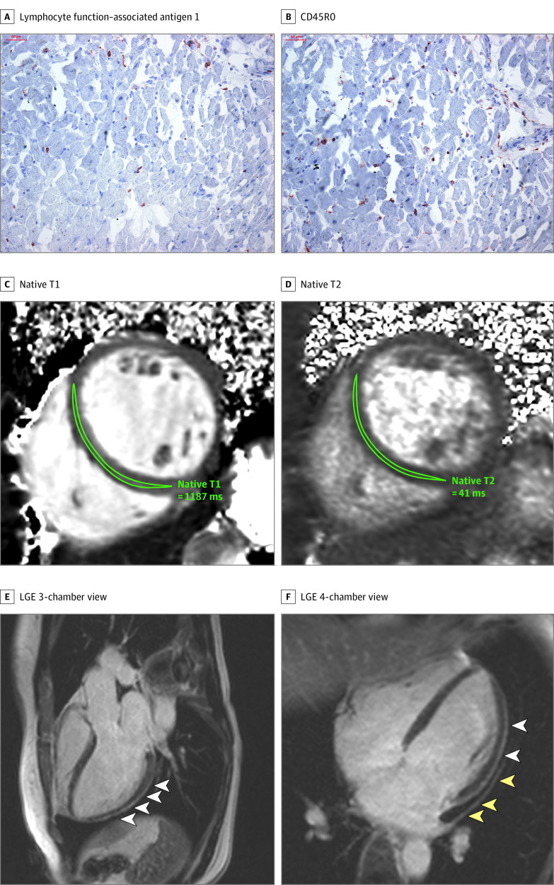
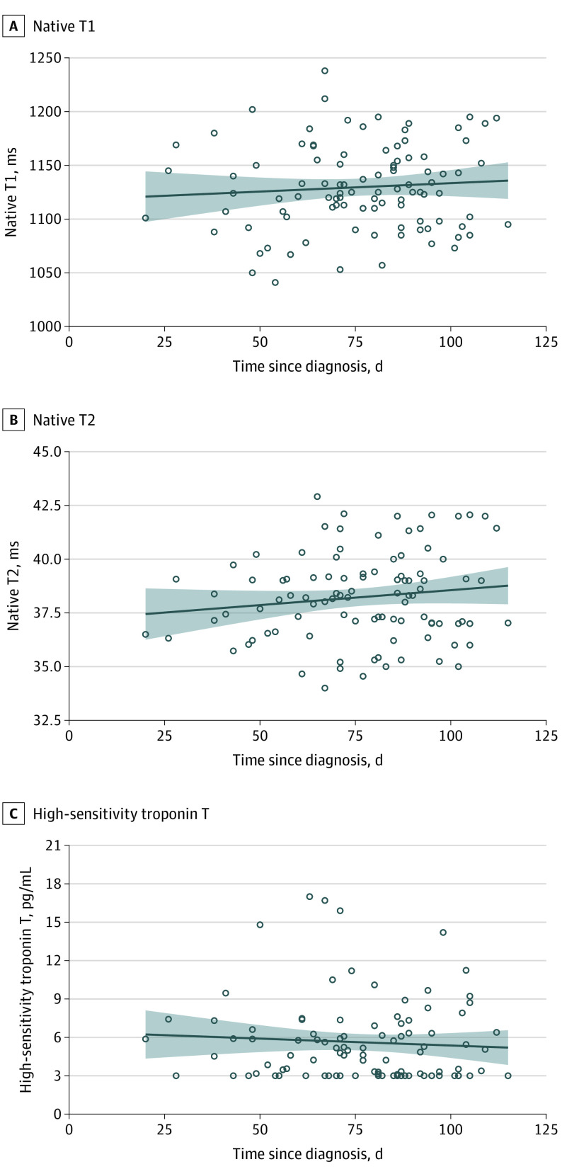
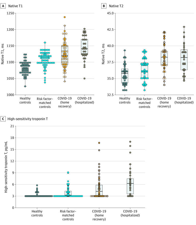
Of the 100 included patients, 53 (53%) were male, and the mean (SD) age was 49 (14) years. The median (IQR) time interval between COVID-19 diagnosis and CMR was 71 (64-92) days. Of the 100 patients recently recovered from COVID-19, 67 (67%) recovered at home, while 33 (33%) required hospitalization. At the time of CMR, high-sensitivity troponin T (hsTnT) was detectable (greater than 3 pg/mL) in 71 patients recently recovered from COVID-19 (71%) and significantly elevated (greater than 13.9 pg/mL) in 5 patients (5%). Compared with healthy controls and risk factor-matched controls, patients recently recovered from COVID-19 had lower left ventricular ejection fraction, higher left ventricle volumes, and raised native T1 and T2. A total of 78 patients recently recovered from COVID-19 (78%) had abnormal CMR findings, including raised myocardial native T1 (n = 73), raised myocardial native T2 (n = 60), myocardial late gadolinium enhancement (n = 32), or pericardial enhancement (n = 22). There was a small but significant difference between patients who recovered at home vs in the hospital for native T1 mapping (median [IQR], 1119 [1092-1150] ms vs 1141 [1121-1175] ms; P = .008) and hsTnT (4.2 [3.0-5.9] pg/dL vs 6.3 [3.4-7.9] pg/dL; P = .002) but not for native T2 mapping. None of these measures were correlated with time from COVID-19 diagnosis (native T1: r = 0.07; P = .47; native T2: r = 0.14; P = .15; hsTnT: r = -0.07; P = .50). High-sensitivity troponin T was significantly correlated with native T1 mapping (r = 0.33; P < .001) and native T2 mapping (r = 0.18; P = .01). Endomyocardial biopsy in patients with severe findings revealed active lymphocytic inflammation. Native T1 and T2 were the measures with the best discriminatory ability to detect COVID-19-related myocardial pathology.

CONCLUSIONS AND RELEVANCE

In this study of a cohort of German patients recently recovered from COVID-19 infection, CMR revealed cardiac involvement in 78 patients (78%) and ongoing myocardial inflammation in 60 patients (60%), independent of preexisting conditions, severity and overall course of the acute illness, and time from the original diagnosis. These findings indicate the need for ongoing investigation of the long-term cardiovascular consequences of COVID-19.

摘要

重要性

2019 年冠状病毒病(COVID-19)继续在全球造成相当大的发病率和死亡率。住院患者的病例报告表明,COVID-19 主要影响心血管系统,但总体影响仍不清楚。

目的

评估最近从 COVID-19 疾病中康复的未选择患者是否存在心肌损伤。

设计、设置和参与者: 在这项前瞻性观察队列研究中,从 2020 年 4 月至 6 月期间法兰克福大学 COVID-19 登记处确定了 100 名最近从 COVID-19 疾病中康复的患者。

暴露

通过上呼吸道拭子检测逆转录-聚合酶链反应确定严重急性呼吸综合征冠状病毒 2 感染的近期康复。

主要结果和测量

获得了人口统计学特征、心脏血液标志物和心血管磁共振(CMR)成像。将其与年龄匹配和性别匹配的健康志愿者对照组(n=50)和风险因素匹配的患者对照组(n=57)进行比较。

结果

在纳入的 100 名患者中,53 名(53%)为男性,平均(SD)年龄为 49(14)岁。COVID-19 诊断与 CMR 之间的中位数(IQR)时间间隔为 71(64-92)天。在 100 名最近从 COVID-19 中康复的患者中,67 名(67%)在家中康复,而 33 名(33%)需要住院治疗。在 CMR 时,71 名(71%)最近从 COVID-19 中康复的患者可检测到高敏肌钙蛋白 T(hsTnT)(大于 3 pg/mL),5 名患者(5%)明显升高(大于 13.9 pg/mL)。与健康对照组和风险因素匹配对照组相比,最近从 COVID-19 中康复的患者左心室射血分数较低,左心室容积较高,心肌固有 T1 和 T2 升高。78 名(78%)最近从 COVID-19 中康复的患者存在异常 CMR 发现,包括升高的心肌固有 T1(n=73)、升高的心肌固有 T2(n=60)、心肌晚期钆增强(n=32)或心包增强(n=22)。在家中康复与在医院康复的患者之间,在心肌固有 T1 图谱(中位数[IQR],1119[1092-1150] ms 与 1141[1121-1175] ms;P=0.008)和 hsTnT(4.2[3.0-5.9] pg/dL 与 6.3[3.4-7.9] pg/dL;P=0.002)方面存在差异,但在心肌固有 T2 图谱方面无差异。这些措施均与 COVID-19 诊断后的时间(固有 T1:r=0.07;P=0.47;固有 T2:r=0.14;P=0.15;hsTnT:r=-0.07;P=0.50)无关。高敏肌钙蛋白 T 与心肌固有 T1 图谱(r=0.33;P<0.001)和心肌固有 T2 图谱(r=0.18;P=0.01)显著相关。在有严重发现的患者中进行的心肌活检显示出活跃的淋巴细胞炎症。固有 T1 和 T2 是检测 COVID-19 相关心肌病理学的最佳鉴别能力的措施。

结论和相关性

在这项对德国最近从 COVID-19 感染中康复的患者队列的研究中,CMR 显示 78 名患者(78%)存在心脏受累,60 名患者(60%)存在持续的心肌炎症,与既往疾病、急性疾病的严重程度和总体病程以及原始诊断后的时间无关。这些发现表明需要进一步研究 COVID-19 的长期心血管后果。

https://cdn.ncbi.nlm.nih.gov/pmc/blobs/4c1d/7385689/57b7e6834496/jamacardiol-e203557-g003.jpg
https://cdn.ncbi.nlm.nih.gov/pmc/blobs/4c1d/7385689/5e9b94c03c52/jamacardiol-e203557-g001.jpg
https://cdn.ncbi.nlm.nih.gov/pmc/blobs/4c1d/7385689/11258b8f5dfa/jamacardiol-e203557-g002.jpg
https://cdn.ncbi.nlm.nih.gov/pmc/blobs/4c1d/7385689/57b7e6834496/jamacardiol-e203557-g003.jpg
https://cdn.ncbi.nlm.nih.gov/pmc/blobs/4c1d/7385689/5e9b94c03c52/jamacardiol-e203557-g001.jpg
https://cdn.ncbi.nlm.nih.gov/pmc/blobs/4c1d/7385689/11258b8f5dfa/jamacardiol-e203557-g002.jpg
https://cdn.ncbi.nlm.nih.gov/pmc/blobs/4c1d/7385689/57b7e6834496/jamacardiol-e203557-g003.jpg

相似文献

1
Outcomes of Cardiovascular Magnetic Resonance Imaging in Patients Recently Recovered From Coronavirus Disease 2019 (COVID-19).COVID-19 患者近期康复后的心血管磁共振成像结果。
JAMA Cardiol. 2020 Nov 1;5(11):1265-1273. doi: 10.1001/jamacardio.2020.3557.
2
Cardiac Involvement in Patients Recovered From COVID-2019 Identified Using Magnetic Resonance Imaging.利用磁共振成像技术识别 COVID-19 康复患者的心脏受累情况。
JACC Cardiovasc Imaging. 2020 Nov;13(11):2330-2339. doi: 10.1016/j.jcmg.2020.05.004. Epub 2020 May 12.
3
T2-mapping increase is the prevalent imaging biomarker of myocardial involvement in active COVID-19: a Cardiovascular Magnetic Resonance study.T2 映射增加是活动性 COVID-19 中心肌受累的主要影像学生物标志物:一项心血管磁共振研究。
J Cardiovasc Magn Reson. 2021 Jun 10;23(1):68. doi: 10.1186/s12968-021-00764-x.
4
Observation of Myocardial Involvement in Patients Recovered From COVID-19 by Using Cardiac Magnetic Resonance Imaging, In a Tertiary Care Hospital, Bangladesh.使用心脏磁共振成像观察孟加拉国一家三级保健医院中 COVID-19 康复患者的心肌受累情况。
Mymensingh Med J. 2022 Oct;31(4):1108-1114.
5
T1 and T2 mapping for evaluation of myocardial involvement in patients with ANCA-associated vasculitides.采用T1和T2映射评估抗中性粒细胞胞浆抗体相关性血管炎患者的心肌受累情况。
J Cardiovasc Magn Reson. 2017 Jan 6;19(1):6. doi: 10.1186/s12968-016-0315-5.
6
Cardiac pathology 6 months after hospitalization for COVID-19 and association with the acute disease severity.COVID-19 住院 6 个月后的心脏病理学变化及其与急性疾病严重程度的关系。
Am Heart J. 2021 Dec;242:61-70. doi: 10.1016/j.ahj.2021.08.001. Epub 2021 Aug 13.
7
Left ventricular function recovery in peripartum cardiomyopathy: a cardiovascular magnetic resonance study by myocardial T1 and T2 mapping.围生期心肌病中心肌 T1 和 T2 mapping 评估左心室功能的恢复:一项心血管磁共振研究。
J Cardiovasc Magn Reson. 2020 Jan 6;22(1):2. doi: 10.1186/s12968-019-0590-z.
8
Cardiac T1 and T2 Mapping Showed Myocardial Involvement in Recovered COVID-19 Patients Initially Considered Devoid of Cardiac Damage.心脏 T1 和 T2 映射显示,最初认为没有心脏损伤的 COVID-19 康复患者存在心肌受累。
J Magn Reson Imaging. 2021 Aug;54(2):421-428. doi: 10.1002/jmri.27534. Epub 2021 Feb 16.
9
Cardiac involvement in COVID-19 patients: mid-term follow up by cardiovascular magnetic resonance.COVID-19 患者的心脏受累:心血管磁共振的中期随访。
J Cardiovasc Magn Reson. 2021 Feb 25;23(1):14. doi: 10.1186/s12968-021-00710-x.
10
Combined Cardiac Fluorodeoxyglucose-Positron Emission Tomography/Magnetic Resonance Imaging Assessment of Myocardial Injury in Patients Who Recently Recovered From COVID-19.近期从 COVID-19 中康复患者的心脏氟脱氧葡萄糖正电子发射断层扫描/磁共振成像心肌损伤的联合评估。
JAMA Cardiol. 2022 Mar 1;7(3):298-308. doi: 10.1001/jamacardio.2021.5505.

引用本文的文献

1
The association between troponin positivity and adverse clinical outcomes, echocardiographic findings, and mortality in hospitalized patients with COVID-19.新冠肺炎住院患者中肌钙蛋白阳性与不良临床结局、超声心动图表现及死亡率之间的关联。
BMC Cardiovasc Disord. 2025 Sep 1;25(1):645. doi: 10.1186/s12872-025-05047-2.
2
Cardio-Pulmonary Features of Long COVID: From Molecular and Histopathological Characteristics to Clinical Implications.长新冠的心肺特征:从分子和组织病理学特征到临床意义
Int J Mol Sci. 2025 Aug 8;26(16):7668. doi: 10.3390/ijms26167668.
3
Implications of Myocardial Fibrosis Burden on Left Ventricular Systolic Function in Sepsis Survivors: Insights from a Retrospective Cohort Study Using Quantitative Late Gadolinium Enhancement Cardiovascular Magnetic Resonance.

本文引用的文献

1
Cardiac Involvement in Patients Recovered From COVID-2019 Identified Using Magnetic Resonance Imaging.利用磁共振成像技术识别 COVID-19 康复患者的心脏受累情况。
JACC Cardiovasc Imaging. 2020 Nov;13(11):2330-2339. doi: 10.1016/j.jcmg.2020.05.004. Epub 2020 May 12.
2
CMR and serology to diagnose COVID-19 infection with primary cardiac involvement.心脏磁共振成像(CMR)和血清学检查用于诊断伴有原发性心脏受累的新型冠状病毒肺炎(COVID-19)感染。
Eur Heart J Cardiovasc Imaging. 2021 Jan 1;22(1):133. doi: 10.1093/ehjci/jeaa169.
3
Diffuse Myocardial Inflammation in COVID-19 Associated Myocarditis Detected by Multiparametric Cardiac Magnetic Resonance Imaging.
脓毒症幸存者中心肌纤维化负担对左心室收缩功能的影响:一项使用定量延迟钆增强心血管磁共振的回顾性队列研究的见解
J Cardiovasc Dev Dis. 2025 Aug 13;12(8):306. doi: 10.3390/jcdd12080306.
4
Low ALT levels and a new diagnosis of congestive heart failure following COVID-19 infection.新冠病毒感染后谷丙转氨酶水平低以及新发充血性心力衰竭诊断
Front Cardiovasc Med. 2025 Aug 5;12:1470239. doi: 10.3389/fcvm.2025.1470239. eCollection 2025.
5
History of COVID-19 as a Risk Factor for Cardiac Arrhythmias: A Case-Control Study.新冠病毒病病史作为心律失常的一个风险因素:一项病例对照研究。
Cardiol Res. 2025 Jun 9;16(4):331-337. doi: 10.14740/cr2042. eCollection 2025 Aug.
6
Long-Term Electrocardiographic Changes in Healthcare Workers Following Mild to Moderate Cases of Coronavirus (COVID-19): A Longitudinal Observational Study.新型冠状病毒(COVID-19)轻症至中症病例后医护人员的长期心电图变化:一项纵向观察研究。
Healthcare (Basel). 2025 Jul 24;13(15):1799. doi: 10.3390/healthcare13151799.
7
Assessment of the myocardial effects of Bradykinin inhibitors by cardiac magnetic resonance (CMR) in discharged COVID-19 patients.在出院的COVID-19患者中,通过心脏磁共振成像(CMR)评估缓激肽抑制剂对心肌的影响。
Sci Rep. 2025 Aug 11;15(1):29340. doi: 10.1038/s41598-025-15489-5.
8
Functional Autoantibodies Targeting G-Protein-Coupled Receptors and Their Clinical Phenotype in Patients with Long-COVID.靶向G蛋白偶联受体的功能性自身抗体及其在长期新冠患者中的临床表型
Int J Mol Sci. 2025 Jul 14;26(14):6746. doi: 10.3390/ijms26146746.
9
Cardiovascular Manifestations of Patients with Long COVID.长期新冠患者的心血管表现
Diagnostics (Basel). 2025 Jul 13;15(14):1771. doi: 10.3390/diagnostics15141771.
10
Influencing factors of early neurological deterioration in COVID-19 -associated acute ischemic stroke.新型冠状病毒肺炎相关急性缺血性卒中早期神经功能恶化的影响因素
Sci Rep. 2025 Jul 27;15(1):27352. doi: 10.1038/s41598-025-13173-2.
多参数心脏磁共振成像检测COVID-19相关性心肌炎中的弥漫性心肌炎症
Circ Cardiovasc Imaging. 2020 May;13(5):e010897. doi: 10.1161/CIRCIMAGING.120.010897. Epub 2020 May 13.
4
Glucocorticoid and immunoglobulin to treat viral fulminant myocarditis.糖皮质激素和免疫球蛋白治疗病毒性暴发性心肌炎。
Eur Heart J. 2020 Jun 7;41(22):2122. doi: 10.1093/eurheartj/ehaa357.
5
Endothelial cell infection and endotheliitis in COVID-19.新型冠状病毒肺炎中的内皮细胞感染与内皮炎
Lancet. 2020 May 2;395(10234):1417-1418. doi: 10.1016/S0140-6736(20)30937-5. Epub 2020 Apr 21.
6
SARS-CoV-2 and viral sepsis: observations and hypotheses.SARS-CoV-2 与病毒败血症:观察与假说。
Lancet. 2020 May 9;395(10235):1517-1520. doi: 10.1016/S0140-6736(20)30920-X. Epub 2020 Apr 17.
7
The ACE2 expression in human heart indicates new potential mechanism of heart injury among patients infected with SARS-CoV-2.人类心脏中的 ACE2 表达表明了 SARS-CoV-2 感染患者心脏损伤的新潜在机制。
Cardiovasc Res. 2020 May 1;116(6):1097-1100. doi: 10.1093/cvr/cvaa078.
8
Cardiac Involvement in a Patient With Coronavirus Disease 2019 (COVID-19).新冠肺炎(COVID-19)患者的心脏受累。
JAMA Cardiol. 2020 Jul 1;5(7):819-824. doi: 10.1001/jamacardio.2020.1096.
9
Cardiovascular Implications of Fatal Outcomes of Patients With Coronavirus Disease 2019 (COVID-19).COVID-19 患者的致命结局对心血管的影响。
JAMA Cardiol. 2020 Jul 1;5(7):811-818. doi: 10.1001/jamacardio.2020.1017.
10
Association of Cardiac Injury With Mortality in Hospitalized Patients With COVID-19 in Wuhan, China.新冠肺炎住院患者中心脏损伤与死亡的相关性研究:中国武汉。
JAMA Cardiol. 2020 Jul 1;5(7):802-810. doi: 10.1001/jamacardio.2020.0950.