Greulich Simon, Mayr Agnes, Kitterer Daniel, Latus Joerg, Henes Joerg, Steubing Hannah, Kaesemann Philipp, Patrascu Alexandru, Greiser Andreas, Groeninger Stefan, Braun Niko, Alscher M Dominik, Sechtem Udo, Mahrholdt Heiko
Division of Cardiology, Robert-Bosch-Medical Center, Auerbachstrasse 110, 70376, Stuttgart, Germany.
Division of Radiology, University Hospital Innsbruck, Innsbruck, Austria.
J Cardiovasc Magn Reson. 2017 Jan 6;19(1):6. doi: 10.1186/s12968-016-0315-5.
Myocardial involvement in AAV patients might be silent, presenting with no or nonspecific symptoms, normal ECG, and preserved left-ventricular ejection fraction (LV-EF). Since up to 50% of deaths in these patients may be due to myocardial involvement, a reliable diagnostic tool is warranted. In contrast to LGE-CMR, which has its strengths in detecting focal inflammatory or fibrotic processes, recent mapping techniques are able to detect even subtle, diffuse inflammatory or fibrotic processes. Our study sought to investigate ANCA (antineutrophil cytoplasmic antibody) associated vasculitides (AAV) patients for myocardial involvement by a cardiovascular magnetic resonance (CMR) protocol, including late gadolinium enhancement (LGE) and mapping sequences.
Thirty seven AAV patients were prospectively enrolled and underwent CMR imaging. Twenty healthy volunteers served as controls.
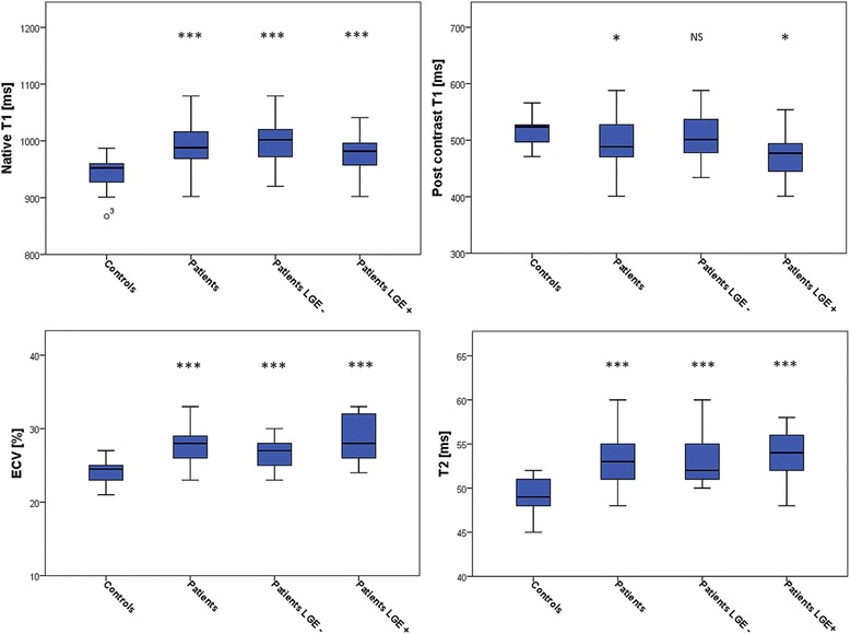
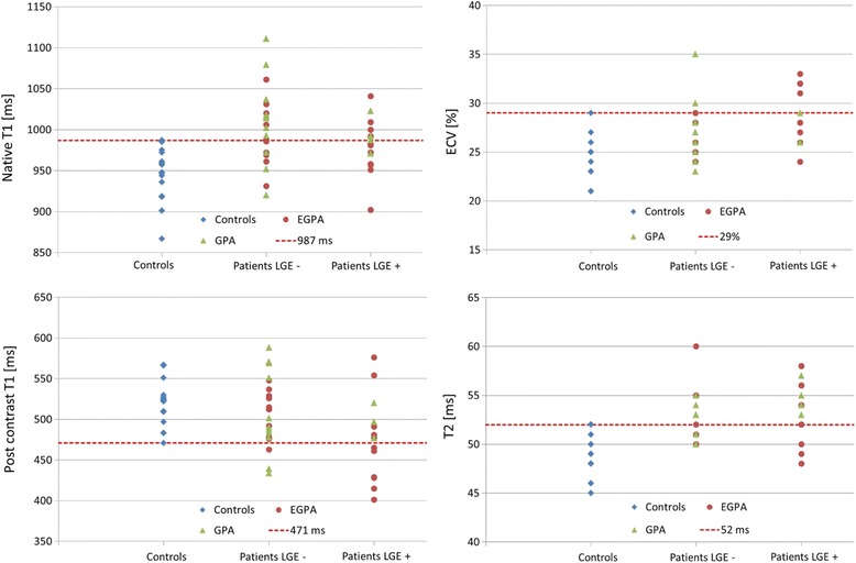
Mean LV-EF was 64%; LGE prevalence of the AAV patients was 43%. AAV patients had higher median native T1 (988 vs. 952 ms, p < 0.001), lower post-contrast T1 (488 vs. 524 ms, p = 0.03), expanded extracellular volume (ECV) (27.5 vs. 24.5%, p < 0.001), and higher T2 (53 vs. 49 ms, p < 0.001) compared to controls, with most parameters independent of the LGE status. Native T1 and T2 in AAV patients showed the highest prevalence of abnormally increased values beyond the 95% percentile of controls.
AAV patients demonstrated increased T1, ECV, and T2 values, with native T1 and T2 showing the highest prevalence of values beyond the 95% percentile of normal. Since these findings seem to be independent of LGE, mapping techniques may provide complementary information to LGE-CMR in the assessment of myocardial involvement in patients with AAV.
AAV患者的心肌受累可能是隐匿性的,表现为无或非特异性症状、心电图正常以及左心室射血分数(LV-EF)保留。由于这些患者中高达50%的死亡可能归因于心肌受累,因此需要一种可靠的诊断工具。与在检测局灶性炎症或纤维化过程方面具有优势的LGE-CMR不同,最近的成像技术能够检测到甚至是细微的、弥漫性的炎症或纤维化过程。我们的研究旨在通过心血管磁共振(CMR)方案,包括延迟钆增强(LGE)和成像序列,来研究抗中性粒细胞胞浆抗体(ANCA)相关血管炎(AAV)患者的心肌受累情况。
前瞻性纳入37例AAV患者并进行CMR成像。20名健康志愿者作为对照。
AAV患者的平均LV-EF为64%;AAV患者的LGE患病率为43%。与对照组相比,AAV患者的中位固有T1更高(988对952毫秒,p<0.001),对比后T1更低(488对524毫秒,p=0.03),细胞外容积(ECV)扩大(27.5对24.5%,p<0.001),T2更高(53对49毫秒,p<0.001),大多数参数与LGE状态无关。AAV患者的固有T1和T2显示,超出对照组95%百分位数的异常升高值患病率最高。
AAV患者表现出T1、ECV和T2值升高,固有T1和T2显示超出正常95%百分位数的值患病率最高。由于这些发现似乎与LGE无关,成像技术可能在评估AAV患者的心肌受累情况时为LGE-CMR提供补充信息。