Galea Nicola, Marchitelli Livia, Pambianchi Giacomo, Catapano Federica, Cundari Giulia, Birtolo Lucia Ilaria, Maestrini Viviana, Mancone Massimo, Fedele Francesco, Catalano Carlo, Francone Marco
Department of Experimental Medicine, "Sapienza" University of Rome, Viale del Policlinico 155, 00161, Rome, Italy.
Department of Radiological, Oncological and Pathological Sciences, "Sapienza" University of Rome, Viale del Policlinico 155, 00161, Rome, Italy.
J Cardiovasc Magn Reson. 2021 Jun 10;23(1):68. doi: 10.1186/s12968-021-00764-x.
Early detection of myocardial involvement can be relevant in coronavirus disease 2019 (COVID-19) patients to timely target symptomatic treatment and decrease the occurrence of the cardiac sequelae of the infection. The aim of the present study was to assess the clinical value of cardiovascular magnetic resonance (CMR) in characterizing myocardial damage in active COVID-19 patients, through the correlation between qualitative and quantitative imaging biomarkers with clinical and laboratory evidence of myocardial injury.
In this retrospective observational cohort study, we enrolled 27 patients with diagnosis of active COVID-19 and suspected cardiac involvement, referred to our institution for CMR between March 2020 and January 2021. Clinical and laboratory characteristics, including high sensitivity troponin T (hs-cTnT), and CMR imaging data were obtained. Relationships between CMR parameters, clinical and laboratory findings were explored. Comparisons were made with age-, sex- and risk factor-matched control group of 27 individuals, including healthy controls and patients without other signs or history of myocardial disease, who underwent CMR examination between January 2020 and January 2021.
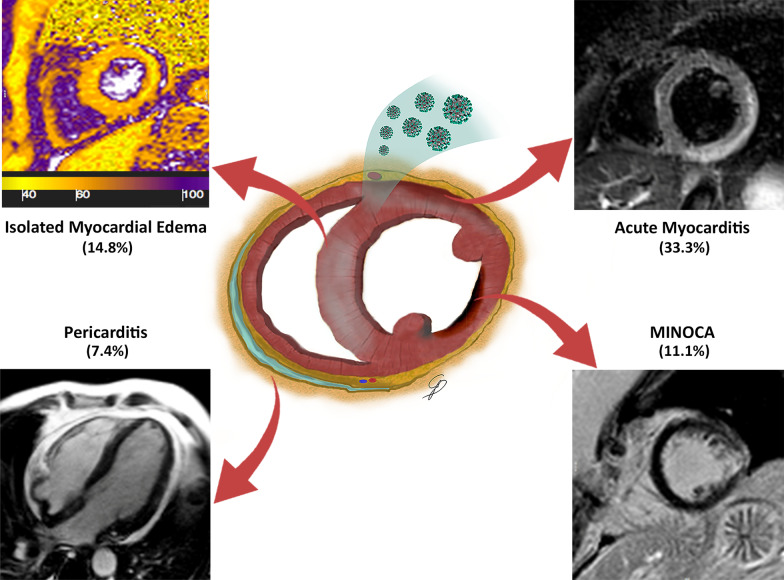
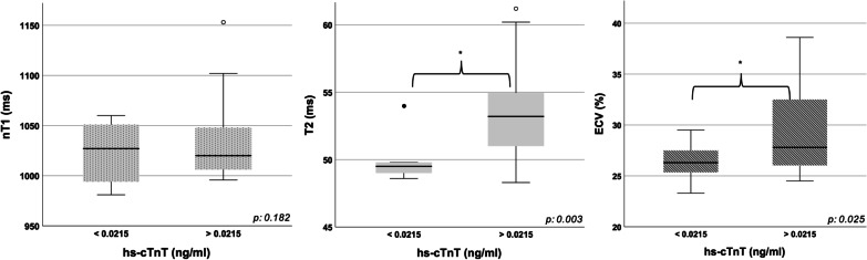
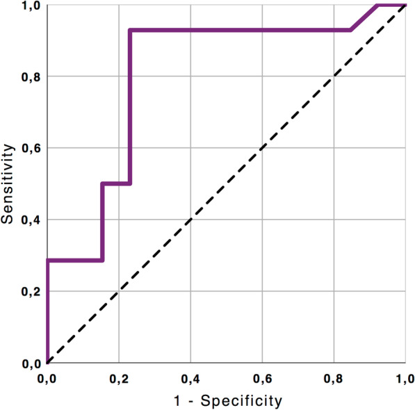
The median (IQR) time interval between COVID-19 diagnosis and CMR examination was 20 (13.5-31.5) days. Hs-cTnT values were collected within 24 h prior to CMR and resulted abnormally increased in 18 patients (66.6%). A total of 20 cases (74%) presented tissue signal abnormalities, including increased myocardial native T1 (n = 11), myocardial T2 (n = 14) and extracellular volume fraction (ECV) (n = 10), late gadolinium enhancement (LGE) (n = 12) or pericardial enhancement (n = 2). A CMR diagnosis of myocarditis was established in 9 (33.3%), pericarditis in 2 (7.4%) and myocardial infarction with non-obstructive coronary arteries in 3 (11.11%) patients. T2 mapping values showed a moderate positive linear correlation with Hs-cTnT (r = 0.58; p = 0.002). A high degree positive linear correlation between ECV and Hs-cTnT was also found (r 0.77; p < 0.001).
CMR allows in vivo recognition and characterization of myocardial damage in a cohort of selected COVID-19 individuals by means of a multiparametric scanning protocol including conventional imaging and T1-T2 mapping sequences. Abnormal T2 mapping was the most commonly abnormality observed in our cohort and positively correlated with hs-cTnT values, reflecting the predominant edematous changes characterizing the active phase of disease.
早期发现心肌受累对于2019冠状病毒病(COVID-19)患者具有重要意义,有助于及时进行对症治疗并减少感染后心脏后遗症的发生。本研究的目的是通过定性和定量成像生物标志物与心肌损伤的临床及实验室证据之间的相关性,评估心血管磁共振(CMR)在表征活动性COVID-19患者心肌损伤方面的临床价值。
在这项回顾性观察队列研究中,我们纳入了27例诊断为活动性COVID-19且怀疑有心脏受累的患者,这些患者于2020年3月至2021年1月被转诊至我院进行CMR检查。获取了临床和实验室特征,包括高敏肌钙蛋白T(hs-cTnT)以及CMR成像数据。探讨了CMR参数、临床和实验室检查结果之间的关系。与27名年龄、性别和危险因素匹配的对照组进行了比较,对照组包括健康对照者以及无其他心肌疾病体征或病史的患者,他们于2020年1月至2021年1月期间接受了CMR检查。
COVID-19诊断与CMR检查之间的中位(IQR)时间间隔为20(13.5 - 31.5)天。hs-cTnT值在CMR检查前24小时内采集,18例患者(66.6%)结果异常升高。共有20例(74%)出现组织信号异常,包括心肌固有T1升高(n = 11)、心肌T2升高(n = 14)和细胞外容积分数(ECV)升高(n = 10)、钆剂延迟强化(LGE)(n = 12)或心包强化(n = 2)。9例(33.3%)患者确诊为心肌炎,2例(7.4%)为心包炎,3例(11.11%)为非阻塞性冠状动脉心肌梗死。T2 mapping值与Hs-cTnT呈中度正线性相关(r = 0.58;p = 0.002)。还发现ECV与Hs-cTnT之间存在高度正线性相关(r = 0.77;p < 0.001)。
CMR能够通过包括传统成像和T1 - T2 mapping序列的多参数扫描方案,在一组选定的COVID-19个体中对心肌损伤进行体内识别和表征。异常的T2 mapping是我们队列中最常见的异常情况,并且与hs-cTnT值呈正相关,反映了疾病活动期主要的水肿变化特征。