Huang Lin, Chen Keliang, Hu Xiaochen, Guo Qihao
Department of Gerontology, Shanghai Jiao Tong University Affiliated Sixth People's Hospital, Shanghai, China.
Department of Neurology, Huashan Hospital, Shanghai Medical College, Fudan University, Shanghai, China.
Front Neurosci. 2020 Jul 9;14:699. doi: 10.3389/fnins.2020.00699. eCollection 2020.
To investigate the bilateral hippocampal subfield volumetric differences in four types of mild dementia, namely typical Alzheimer's disease (tAD), dementia with Lewy bodies (DLB), semantic dementia (SD), and posterior cortical atrophy (PCA), to assist differential diagnosis.
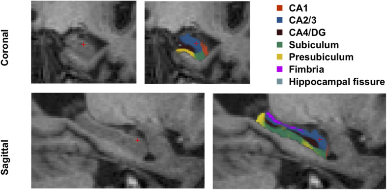
One hundred three participants, including 22 tAD, 34 SD (17 left SD and 17 right SD), 15 DLB, 12 PCA patients, and 20 normal controls (NC), were recruited. All subjects received standard neuropsychological assessments and magnetic resonance imaging (MRI). The hippocampal subfields were automatically segmented via Freesurfer. The study compared the volumetric differences and used the receiver operating characteristic (ROC) curves to estimate the efficacy of each hippocampal subfield to distinguish between groups. Spearman correlation analysis was used to investigate the relationship between memory recall scores and hippocampal subfield volumes.
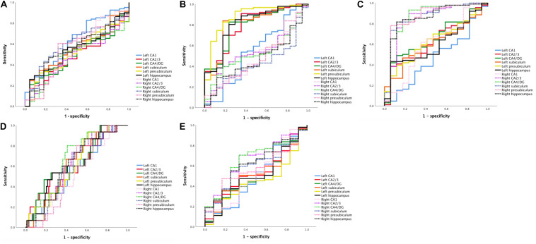
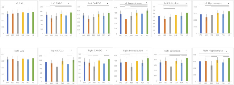
The hippocampal subfield atrophy varied in different groups: tAD, SD, and PCA patients had subregional atrophy in bilateral hippocampi compared to NC, and DLB patients showed preserved volumes; left SD patients suffered the most severe atrophy of the left hippocampus, and right SD patients were atrophied mostly in the right hippocampus. There was no significant difference in the volume of hippocampal subregions between tAD and PCA subjects, but the former tended to be atrophied more asymmetrically. ROC analysis showed that, for discrimination, the areas under the curve (AUC) of some subfields were larger than the total hippocampus, but none observed significant difference. In addition, immediate recall scores were correlated to left CA1, CA2/3, CA4/DG, subiculum, and presubiculum ( < 0.05), and delayed recall scores were strongly related to bilateral CA2/3, CA4/DG, subiculum, and presubiculum ( = 0.38-0.52, < 0.05).
Differential atrophy patterns in the bilateral hippocampal subfield volumes could serve the differential diagnosis in patients with different causes of mild dementia: left CA1 for tAD; left presubiculum for LSD; right CA4/DG, right presubiculum, and right subiculum for RSD; CA4/DG and right CA2/3 for DLB; right CA2/3 and right CA4/DG for PCA. Additionally, several hippocampal subfield volumes were significantly associated with memory scores, further highlighting the essential role of the hippocampus in memory decline.
研究四种轻度痴呆类型,即典型阿尔茨海默病(tAD)、路易体痴呆(DLB)、语义性痴呆(SD)和后部皮质萎缩(PCA)的双侧海马亚区体积差异,以辅助鉴别诊断。
招募了103名参与者,包括22名tAD患者、34名SD患者(17名左侧SD和17名右侧SD)、15名DLB患者、12名PCA患者以及20名正常对照(NC)。所有受试者均接受了标准神经心理学评估和磁共振成像(MRI)检查。通过Freesurfer自动分割海马亚区。本研究比较了体积差异,并使用受试者工作特征(ROC)曲线来评估每个海马亚区区分不同组别的效能。采用Spearman相关性分析来研究记忆回忆分数与海马亚区体积之间的关系。
不同组别的海马亚区萎缩情况各不相同:与NC相比,tAD、SD和PCA患者双侧海马均存在亚区域萎缩,而DLB患者的体积保持正常;左侧SD患者左侧海马萎缩最为严重,右侧SD患者右侧海马萎缩最为明显。tAD和PCA受试者之间海马亚区的体积没有显著差异,但前者往往萎缩更不对称。ROC分析表明,为了进行鉴别,一些亚区的曲线下面积(AUC)大于整个海马,但均未观察到显著差异。此外,即时回忆分数与左侧CA1、CA2/3、CA4/DG、下托和前下托相关(<0.05),延迟回忆分数与双侧CA2/3、CA4/DG、下托和前下托密切相关(=0.38 - 0.52,<0.05)。
双侧海马亚区体积的差异萎缩模式可用于不同病因轻度痴呆患者的鉴别诊断:左侧CA1用于tAD;左侧前下托用于左侧SD;右侧CA4/DG、右侧前下托和右侧下托用于右侧SD;CA4/DG和右侧CA2/3用于DLB;右侧CA2/3和右侧CA4/DG用于PCA。此外,几个海马亚区的体积与记忆分数显著相关,进一步突出了海马在记忆衰退中的重要作用。