de Krijger Manon, Visseren Thijmen, Wildenberg Manon E, Hooijer Gerrit K J, Verstegen Monique M A, van der Laan Luc J W, de Jonge Wouter J, Verheij Joanne, Ponsioen Cyriel Y
Tytgat Institute for Liver and Intestinal Research, Amsterdam UMC, University of Amsterdam, Meibergdreef 69, 1105 BK, Amsterdam, the Netherlands.
Department of Gastroenterology and Hepatology Amsterdam UMC, University of Amsterdam, Meibergdreef 9, 1105 AZ, Amsterdam, the Netherlands.
J Transl Autoimmun. 2020 Apr 9;3:100054. doi: 10.1016/j.jtauto.2020.100054. eCollection 2020.
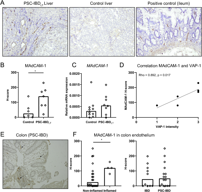
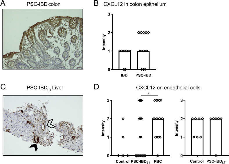
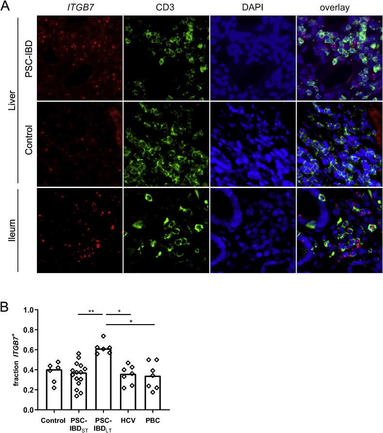
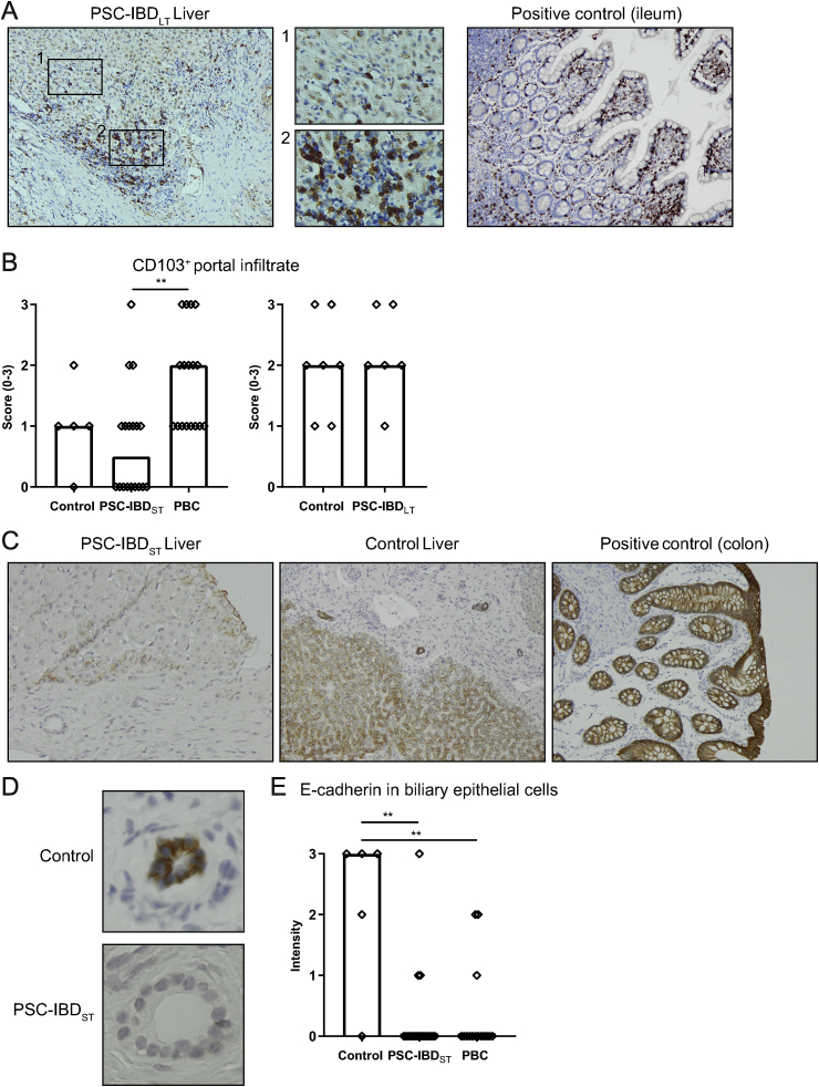
The co-occurrence of inflammatory bowel disease (IBD) in up to 80% of patients with primary sclerosing cholangitis (PSC) suggests a relation between the gut and the liver in patients with both PSC and IBD. One hypothesis suggests that aberrantly expressed homing molecules in the liver drive infiltration of gut-homing memory T-cells that are originally primed in intestinal environment. One of the main findings supporting this hypothesis is the expression of mucosal addressin cell adhesion molecule 1 (MAdCAM-1) in PSC livers. Expression of homing molecules in early PSC remains unclear. The aim of this study was to investigate expression patterns of homing chemokines and adhesion molecules in PSC-IBD colons and livers, and to study whether changes are already present in early stages of PSC.
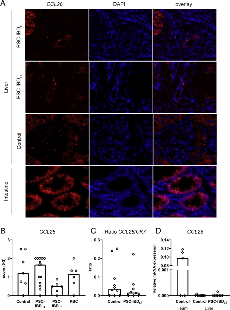
Needle biopsies from livers of 20 PSC patients with short-term PSC (PSC-IBD) as well as explant liver biopsies of 8 patients with long-term PSC (PSC-IBD) were collected (median disease duration 0 and 22 years, respectively). Only patients with concomitant IBD were included (89% ulcerative colitis and 11% Crohn's disease). Expression and distribution of MAdCAM-1, VAP-1, integrin β7, CCL25, CCL28, CXCL12, αE (CD103) and E-cadherin were assessed in both liver and colon tissue. Liver tissue collected from obstructive cholangitis in resection specimens for Klatskin tumors or resection specimens from hepatic metastasis, liver tissue of patients with hepatitis C virus (HCV) and of patients with primary biliary cholangitis (PBC) served as controls.
MAdCAM-1 expression in livers of PSC-IBD patients was increased compared to controls. The proportion of CD3 T-cells expressing integrin β7 did not differ between PSC-IBD and control groups, but was higher in liver tissue of PSC-IBD patients. There was no difference in αE T-cells between PSC-IBD and control groups. The chemokine CCL28 was highly expressed in biliary epithelial cells. This intense staining pattern was more pronounced in PSC-IBD, but overall did not significantly differ from controls.
We confirm that aberrant gut lymphocyte homing to the liver exists in PSC, linking gut and liver disease pathology in PSC-IBD. Our data suggests that this phenomenon increases over time in later stages of the disease, worsening ongoing inflammation.
高达80%的原发性硬化性胆管炎(PSC)患者同时患有炎症性肠病(IBD),这表明PSC和IBD患者的肠道与肝脏之间存在关联。一种假说认为,肝脏中异常表达的归巢分子促使原本在肠道环境中致敏的肠道归巢记忆T细胞浸润。支持这一假说的主要发现之一是PSC肝脏中黏膜地址素细胞黏附分子1(MAdCAM-1)的表达。早期PSC中归巢分子的表达尚不清楚。本研究的目的是调查PSC-IBD结肠和肝脏中归巢趋化因子和黏附分子的表达模式,并研究PSC早期是否已出现变化。
收集20例短期PSC患者(PSC-IBD)肝脏的针吸活检组织以及8例长期PSC患者(PSC-IBD)的肝外植体活检组织(疾病持续时间中位数分别为0年和22年)。仅纳入合并IBD的患者(89%为溃疡性结肠炎,11%为克罗恩病)。评估MAdCAM-1、VAP-1、整合素β7、CCL25、CCL28、CXCL12、αE(CD103)和E-钙黏蛋白在肝脏和结肠组织中的表达及分布。从肝门部胆管癌切除标本或肝转移切除标本中的阻塞性胆管炎收集的肝脏组织、丙型肝炎病毒(HCV)患者及原发性胆汁性胆管炎(PBC)患者的肝脏组织作为对照。
与对照组相比,PSC-IBD患者肝脏中MAdCAM-1的表达增加。表达整合素β7的CD3 T细胞比例在PSC-IBD组和对照组之间无差异,但在PSC-IBD患者的肝脏组织中更高。PSC-IBD组和对照组之间αE T细胞无差异。趋化因子CCL28在胆管上皮细胞中高表达。这种强烈的染色模式在PSC-IBD中更明显,但总体上与对照组无显著差异。
我们证实PSC中存在异常的肠道淋巴细胞向肝脏归巢,这将PSC-IBD中的肠道和肝脏疾病病理联系起来。我们的数据表明,这种现象在疾病后期会随着时间增加,加剧持续的炎症。