Department of Radiological, Oncological and Pathological Sciences, Policlinico Umberto I, Sapienza University of Rome, Italy.
Department of Emergency and Acceptance, Anesthesiology and Intensive Care Unit, Policlinico Umberto I, Sapienza University of Rome, Italy.
Eur J Radiol. 2020 Sep;130:109202. doi: 10.1016/j.ejrad.2020.109202. Epub 2020 Jul 29.
So far, only a few studies evaluated the correlation between CT features and clinical outcome in patients with COVID-19 pneumonia.
To evaluate CT ability in differentiating critically ill patients requiring invasive ventilation from patients with less severe disease.
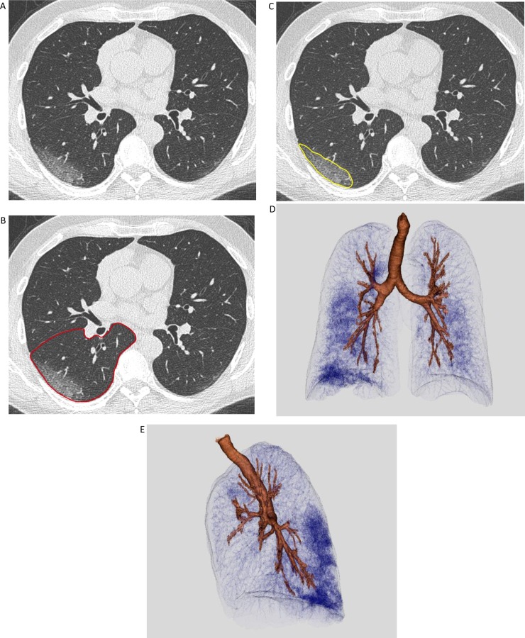
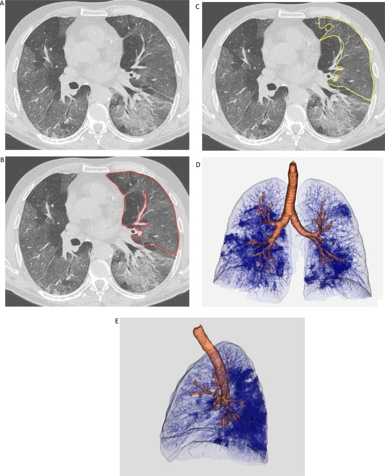
We retrospectively collected data from patients admitted to our institution for COVID-19 pneumonia between March 5th-24th. Patients were considered critically ill or non-critically ill, depending on the need for mechanical ventilation. CT images from both groups were analyzed for the assessment of qualitative features and disease extension, using a quantitative semiautomatic method. We evaluated the differences between the two groups for clinical, laboratory and CT data. Analyses were conducted on a per-protocol basis.
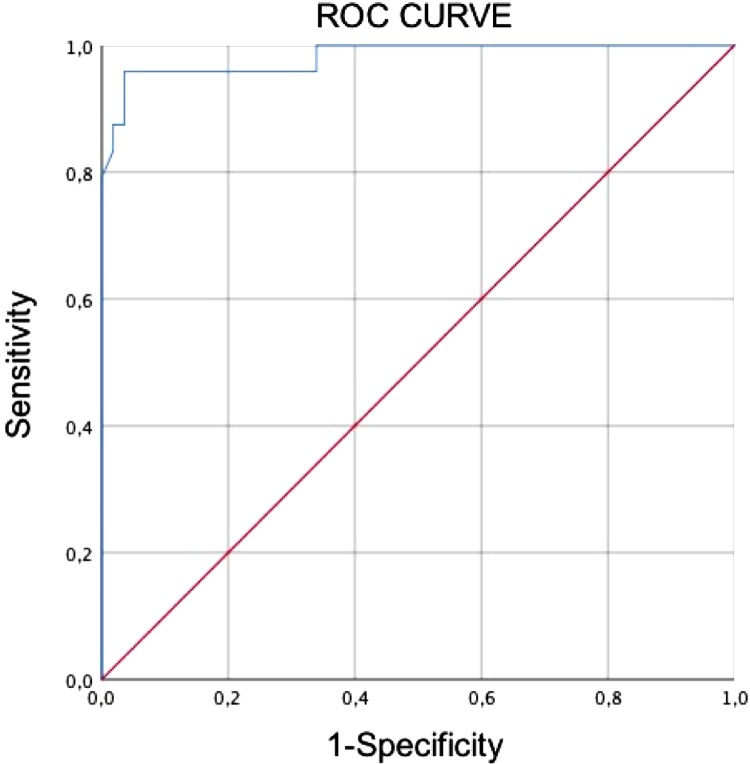
189 patients were analyzed. PaO/FIO ratio and oxygen saturation (SaO) were decreased in critically ill patients. At CT, mixed pattern (ground glass opacities (GGO) and consolidation) and GGO alone were more frequent respectively in critically ill and in non-critically ill patients (p < 0.05). Lung volume involvement was significantly higher in critically ill patients (38.5 % vs. 5.8 %, p < 0.05). A cut-off of 23.0 % of lung involvement showed 96 % sensitivity and 96 % specificity in distinguishing critically ill patients from patients with less severe disease. The fraction of involved lung was related to lactate dehydrogenase (LDH) levels, PaO/FIO ratio and SaO (p < 0.05).
Lung disease extension, assessed using quantitative CT, has a significant relationship with clinical severity and may predict the need for invasive ventilation in patients with COVID-19.
目前,仅有少数研究评估了 COVID-19 肺炎患者的 CT 特征与临床结局之间的相关性。
评估 CT 区分需要有创通气的危重症患者与病情较轻患者的能力。
我们回顾性收集了 3 月 5 日至 24 日期间因 COVID-19 肺炎入住我院的患者数据。根据是否需要机械通气,将患者分为危重症或非危重症。使用定量半自动方法分析两组 CT 图像,评估定性特征和疾病进展。我们评估了两组间的临床、实验室和 CT 数据差异。分析基于意向治疗原则。
共分析了 189 例患者。危重症患者的 PaO/FIO 比值和氧饱和度(SaO)降低。在 CT 上,磨玻璃影(GGO)和实变混合模式以及单纯 GGO 在危重症和非危重症患者中更为常见(p<0.05)。危重症患者的肺容积受累明显更高(38.5% vs. 5.8%,p<0.05)。肺受累 23.0%的截断值在区分危重症患者和病情较轻患者方面具有 96%的敏感性和 96%的特异性。受累肺的比例与乳酸脱氢酶(LDH)水平、PaO/FIO 比值和 SaO 相关(p<0.05)。
使用定量 CT 评估的肺部疾病进展与临床严重程度具有显著相关性,可能预测 COVID-19 患者是否需要有创通气。