• 文献检索
  • 文档翻译
  • 深度研究
  • 学术资讯
  • Suppr Zotero 插件Zotero 插件
  • 邀请有礼
  • 套餐&价格
  • 历史记录
应用&插件
Suppr Zotero 插件Zotero 插件浏览器插件Mac 客户端Windows 客户端微信小程序
定价
高级版会员购买积分包购买API积分包
服务
文献检索文档翻译深度研究API 文档MCP 服务
关于我们
关于 Suppr公司介绍联系我们用户协议隐私条款
关注我们

Suppr 超能文献

核心技术专利:CN118964589B侵权必究
粤ICP备2023148730 号-1Suppr @ 2026

文献检索

告别复杂PubMed语法,用中文像聊天一样搜索,搜遍4000万医学文献。AI智能推荐,让科研检索更轻松。

立即免费搜索

文件翻译

保留排版,准确专业,支持PDF/Word/PPT等文件格式,支持 12+语言互译。

免费翻译文档

深度研究

AI帮你快速写综述,25分钟生成高质量综述,智能提取关键信息,辅助科研写作。

立即免费体验

以症状持续时间为特征的 COVID-19 肺炎的胸部 CT 表现。

Chest CT findings of COVID-19 pneumonia by duration of symptoms.

机构信息

Department of Radiology, Zhongnan Hospital of Wuhan University, Wuhan University, Wuhan, 430071, PR China.

出版信息

Eur J Radiol. 2020 Jun;127:109009. doi: 10.1016/j.ejrad.2020.109009. Epub 2020 Apr 18.

DOI:10.1016/j.ejrad.2020.109009
PMID:32325282
原文链接:https://pmc.ncbi.nlm.nih.gov/articles/PMC7165099/
Abstract

PURPOSE

To evaluate lung abnormalities on thin-section computed tomographic (CT) scans in patients with COVID-19 and correlate findings to duration of symptoms.

METHODS

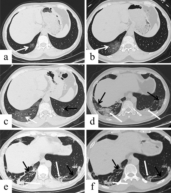
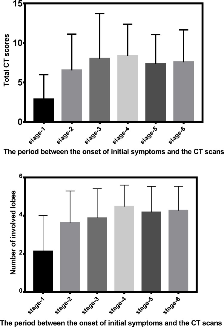
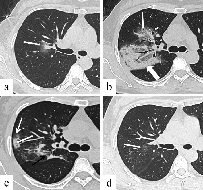
In total, 348 CT scans in 112 patients were classified according to the time after the onset of the initial symptoms, namely stage-1 (0-4 days); stage-2 (5-9 days); stage-3 (10-14 days); stage-4 (15-21 days); stage-5 (22-28 days); and stage-6 (>28 days). Each lung lobe was evaluated for extent affected by ground-glass opacities (GGO), crazy-paving pattern and consolidation, in five categories of percentual severity. Summation of scores from all five lung lobes provided the total CT score (maximal CT score, 25).

RESULTS

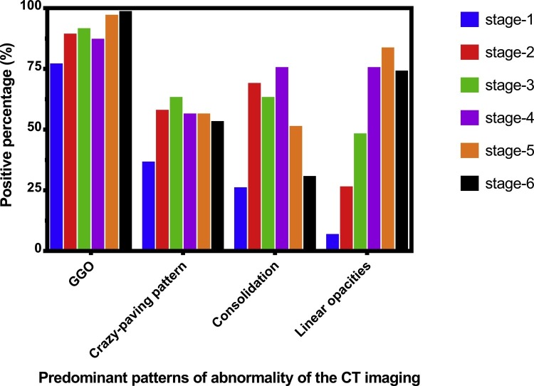
The predominant patterns of lung abnormalities were GGOs, crazy-paving pattern, consolidation and linear opacities. The frequency of crazy-paving pattern, consolidation and linear opacities peaked at stage-3 (62.7 %), stage-4 (75.0 %) and stage-5 (83.1 %), respectively, and decreased thereafter. Total CT scores increased from stage-1 to stage-2 (2.8 ± 3.1, vs. 6.5 ± 4.6, respectively, P < 0.01), and thereafter remained high. The lower lobes were more inclined to be involved with higher CT scores except for stage-1. At stage-6 98.1 % of CT scans still showed abnormalities (CT score 7.5 ± 4.1).

CONCLUSION

Thin-section CT could provide semi-quantitative analysis of pulmonary damage severity. This disease changed rapidly at the early stage, then tended to be stable and lasted for a long time.

摘要

目的

评估 COVID-19 患者的肺部 CT 扫描的异常情况,并将其与症状持续时间相关联。

方法

总共对 112 名患者的 348 个 CT 扫描进行分类,根据初始症状发作后的时间,即第 1 期(0-4 天);第 2 期(5-9 天);第 3 期(10-14 天);第 4 期(15-21 天);第 5 期(22-28 天);第 6 期(>28 天)。每个肺叶均按磨玻璃影(GGO)、铺路石征和实变的程度分为五个等级进行评估。五个肺叶评分总和即为总 CT 评分(最大 CT 评分,25 分)。

结果

肺部异常的主要表现为 GGO、铺路石征、实变和线性混浊。铺路石征、实变和线性混浊的频率在第 3 期(62.7%)、第 4 期(75.0%)和第 5 期(83.1%)达到峰值,此后逐渐下降。总 CT 评分从第 1 期到第 2 期(分别为 2.8±3.1 分和 6.5±4.6 分,P<0.01)升高,此后保持较高水平。除第 1 期外,下叶更容易出现较高的 CT 评分。在第 6 期,98.1%的 CT 扫描仍显示异常(CT 评分 7.5±4.1)。

结论

薄层 CT 可对肺部损伤严重程度进行半定量分析。这种疾病在早期变化迅速,随后趋于稳定并持续很长时间。

https://cdn.ncbi.nlm.nih.gov/pmc/blobs/679a/7165099/6f2409ff3033/gr4_lrg.jpg
https://cdn.ncbi.nlm.nih.gov/pmc/blobs/679a/7165099/eced70f93d91/gr1_lrg.jpg
https://cdn.ncbi.nlm.nih.gov/pmc/blobs/679a/7165099/cad838586eb4/gr2_lrg.jpg
https://cdn.ncbi.nlm.nih.gov/pmc/blobs/679a/7165099/fe0d21ebf104/gr3_lrg.jpg
https://cdn.ncbi.nlm.nih.gov/pmc/blobs/679a/7165099/6f2409ff3033/gr4_lrg.jpg

相似文献

1
Chest CT findings of COVID-19 pneumonia by duration of symptoms.以症状持续时间为特征的 COVID-19 肺炎的胸部 CT 表现。
Eur J Radiol. 2020 Jun;127:109009. doi: 10.1016/j.ejrad.2020.109009. Epub 2020 Apr 18.
2
Time Course of Lung Changes at Chest CT during Recovery from Coronavirus Disease 2019 (COVID-19).新冠肺炎(COVID-19)康复过程中胸部 CT 肺部变化的时间进程。
Radiology. 2020 Jun;295(3):715-721. doi: 10.1148/radiol.2020200370. Epub 2020 Feb 13.
3
A Comparison of Clinical and Chest CT Findings in Patients With Influenza A (H1N1) Virus Infection and Coronavirus Disease (COVID-19).甲型流感病毒(H1N1)感染与新型冠状病毒肺炎(COVID-19)患者的临床和胸部 CT 表现比较。
AJR Am J Roentgenol. 2020 Nov;215(5):1065-1071. doi: 10.2214/AJR.20.23214. Epub 2020 May 26.
4
Coronavirus disease 2019: initial chest CT findings.新型冠状病毒肺炎的胸部 CT 表现。
Eur Radiol. 2020 Aug;30(8):4398-4406. doi: 10.1007/s00330-020-06816-7. Epub 2020 Mar 24.
5
[Chest CT findings and their dynamic changes in patients with COVID-19].新型冠状病毒肺炎患者胸部CT表现及其动态变化
Nan Fang Yi Ke Da Xue Xue Bao. 2020 Mar 30;40(3):327-332. doi: 10.12122/j.issn.1673-4254.2020.03.05.
6
Evolution of CT findings in patients with mild COVID-19 pneumonia.轻度 COVID-19 肺炎患者的 CT 表现演变。
Eur Radiol. 2020 Sep;30(9):4865-4873. doi: 10.1007/s00330-020-06823-8. Epub 2020 Apr 15.
7
CT in coronavirus disease 2019 (COVID-19): a systematic review of chest CT findings in 4410 adult patients.CT 在 2019 年冠状病毒病(COVID-19)中的应用:对 4410 例成年患者胸部 CT 表现的系统综述。
Eur Radiol. 2020 Nov;30(11):6129-6138. doi: 10.1007/s00330-020-06975-7. Epub 2020 May 30.
8
Spectrum of chest computed tomographic (CT) findings in coronavirus disease-19 (COVID-19) patients in India.印度 COVID-19 患者的胸部计算机断层扫描(CT)表现谱。
Eur J Radiol. 2020 Aug;129:109147. doi: 10.1016/j.ejrad.2020.109147. Epub 2020 Jun 24.
9
CT findings of COVID-19 in follow-up: comparison between progression and recovery.新冠肺炎 CT 随访表现:进展与恢复的比较。
Diagn Interv Radiol. 2020 Jul;26(4):301-307. doi: 10.5152/dir.2019.20176.
10
Imaging and clinical features of patients with 2019 novel coronavirus SARS-CoV-2.新型冠状病毒 2019 年 SARS-CoV-2 患者的影像学和临床特征。
Eur J Nucl Med Mol Imaging. 2020 May;47(5):1275-1280. doi: 10.1007/s00259-020-04735-9. Epub 2020 Feb 28.

引用本文的文献

1
Defining the Subtypes of Long COVID and Risk Factors for Prolonged Disease: Population-Based Case-Crossover Study.定义长新冠的亚型和疾病迁延的危险因素:基于人群的病例交叉研究。
JMIR Public Health Surveill. 2024 Apr 30;10:e49841. doi: 10.2196/49841.
2
Retrospective computed tomography assessment of chemotherapy-related pneumonia with severity screening in pediatric acute lymphoblastic leukemia by radiological imaging.通过放射影像学对小儿急性淋巴细胞白血病化疗相关肺炎进行严重程度筛查的回顾性计算机断层扫描评估
Heliyon. 2023 Dec 9;10(1):e23444. doi: 10.1016/j.heliyon.2023.e23444. eCollection 2024 Jan 15.
3
Comparison of Chest Computed Tomography (CT) Imaging Patterns and Severity among COVID-19 Patients during the First and Fourth Waves in Addis Ababa, Ethiopia.

本文引用的文献

1
Asymptomatic SARS-CoV-2 infected patients with persistent negative CT findings.CT检查结果持续呈阴性的无症状新型冠状病毒感染患者。
Eur J Radiol. 2020 May;126:108956. doi: 10.1016/j.ejrad.2020.108956. Epub 2020 Mar 12.
2
Experience of different upper respiratory tract sampling strategies for detection of COVID-19.不同上呼吸道采样策略用于检测新型冠状病毒肺炎的经验
J Hosp Infect. 2020 May;105(1):1-2. doi: 10.1016/j.jhin.2020.03.012. Epub 2020 Mar 12.
3
Sensitivity of Chest CT for COVID-19: Comparison to RT-PCR.胸部CT对新型冠状病毒肺炎的敏感性:与逆转录聚合酶链反应的比较。
埃塞俄比亚亚的斯亚贝巴新冠疫情第一波和第四波期间新冠肺炎患者胸部计算机断层扫描(CT)成像模式与严重程度的比较
Radiol Res Pract. 2023 Nov 29;2023:6385162. doi: 10.1155/2023/6385162. eCollection 2023.
4
Radiological Patterns of Lung Injury on HRCT in COVID-19 Patients: An Experience From Tertiary Care Centre in India.新型冠状病毒肺炎患者高分辨率CT上肺损伤的影像学表现:来自印度一家三级医疗中心的经验
Cureus. 2023 Nov 7;15(11):e48479. doi: 10.7759/cureus.48479. eCollection 2023 Nov.
5
Sialylated Glycan Bindings from SARS-CoV-2 Spike Protein to Blood and Endothelial Cells Govern the Severe Morbidities of COVID-19.SARS-CoV-2 刺突蛋白与血液和血管内皮细胞的唾液酸化聚糖结合决定了 COVID-19 的严重发病机制。
Int J Mol Sci. 2023 Dec 1;24(23):17039. doi: 10.3390/ijms242317039.
6
Effects of pulmonary air leak on patients with coronavirus disease 2019 (COVID-19): a systematic review and meta-analysis.肺空气漏对 2019 冠状病毒病(COVID-19)患者的影响:系统评价和荟萃分析。
BMC Pulm Med. 2023 Oct 19;23(1):398. doi: 10.1186/s12890-023-02710-2.
7
Evaluation of computed tomography images under deep learning in the diagnosis of severe pulmonary infection.深度学习下计算机断层扫描图像在重症肺部感染诊断中的评估
Front Comput Neurosci. 2023 Aug 4;17:1115167. doi: 10.3389/fncom.2023.1115167. eCollection 2023.
8
Investigating of the role of CT scan for cancer patients during the first wave of COVID-19 pandemic.在新冠疫情第一波期间对癌症患者进行CT扫描作用的调查。
Res Diagn Interv Imaging. 2022 Mar;1:100004. doi: 10.1016/j.redii.2022.100004. Epub 2022 Mar 31.
9
Parasternal intercostal thickening at hospital admission: a promising indicator for mechanical ventilation risk in subjects with severe COVID-19.入院时胸骨旁肋间增厚:严重 COVID-19 患者机械通气风险的有前途指标。
J Clin Monit Comput. 2023 Oct;37(5):1287-1293. doi: 10.1007/s10877-023-00989-4. Epub 2023 Mar 24.
10
Repeated Lung Ultrasound versus Chest X-ray-Which One Predicts Better Clinical Outcome in COVID-19?重复肺部超声与胸部 X 线片——哪一种能更好地预测 COVID-19 的临床结局?
Tomography. 2023 Mar 21;9(2):706-716. doi: 10.3390/tomography9020056.
Radiology. 2020 Aug;296(2):E115-E117. doi: 10.1148/radiol.2020200432. Epub 2020 Feb 19.
4
Initial CT findings and temporal changes in patients with the novel coronavirus pneumonia (2019-nCoV): a study of 63 patients in Wuhan, China.新型冠状病毒肺炎(2019-nCoV)患者的初始 CT 表现及时间演变:中国武汉 63 例患者研究。
Eur Radiol. 2020 Jun;30(6):3306-3309. doi: 10.1007/s00330-020-06731-x. Epub 2020 Feb 13.
5
Time Course of Lung Changes at Chest CT during Recovery from Coronavirus Disease 2019 (COVID-19).新冠肺炎(COVID-19)康复过程中胸部 CT 肺部变化的时间进程。
Radiology. 2020 Jun;295(3):715-721. doi: 10.1148/radiol.2020200370. Epub 2020 Feb 13.
6
Chest CT for Typical Coronavirus Disease 2019 (COVID-19) Pneumonia: Relationship to Negative RT-PCR Testing.胸部 CT 与典型 2019 冠状病毒病(COVID-19)肺炎:与阴性 RT-PCR 检测的关系。
Radiology. 2020 Aug;296(2):E41-E45. doi: 10.1148/radiol.2020200343. Epub 2020 Feb 12.
7
Public responses to the novel 2019 coronavirus (2019-nCoV) in Japan: Mental health consequences and target populations.日本公众对新型2019冠状病毒(2019-nCoV)的反应:心理健康影响及目标人群。
Psychiatry Clin Neurosci. 2020 Apr;74(4):281-282. doi: 10.1111/pcn.12988. Epub 2020 Feb 23.
8
Detection of 2019 novel coronavirus (2019-nCoV) by real-time RT-PCR.实时 RT-PCR 检测 2019 新型冠状病毒(2019-nCoV)
Euro Surveill. 2020 Jan;25(3). doi: 10.2807/1560-7917.ES.2020.25.3.2000045.
9
Clinical features of patients infected with 2019 novel coronavirus in Wuhan, China.中国武汉地区 2019 年新型冠状病毒感染患者的临床特征。
Lancet. 2020 Feb 15;395(10223):497-506. doi: 10.1016/S0140-6736(20)30183-5. Epub 2020 Jan 24.
10
Pulmonary sequelae in convalescent patients after severe acute respiratory syndrome: evaluation with thin-section CT.严重急性呼吸综合征康复患者的肺部后遗症:薄层CT评估
Radiology. 2005 Sep;236(3):1067-75. doi: 10.1148/radiol.2363040958. Epub 2005 Jul 29.