Epilepsy Society MRI Unit, Chalfont Centre for Epilepsy, Chalfont St Peter, SL9 0LR, UK.
Department of Clinical and Experimental Epilepsy, UCL Queen Square Institute of Neurology, Queen Square, London, WC1N 3BG, UK.
J Neurol. 2021 Jan;268(1):147-160. doi: 10.1007/s00415-020-10116-x. Epub 2020 Aug 3.
To investigate alterations of language networks and their relation to impaired naming performance in temporal lobe epilepsy (TLE) using functional MRI.
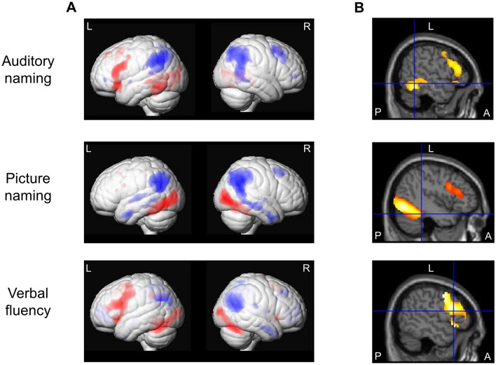
Seventy-two adult TLE patients (41 left) and 36 controls were studied with overt auditory and picture naming fMRI tasks to assess temporal lobe language areas, and a covert verbal fluency task to probe frontal lobe language regions. Correlation of fMRI activation with clinical naming scores, and alteration of language network patterns in relation to epilepsy duration, age at onset and seizure frequency, were investigated with whole-brain multiple regression analyses.
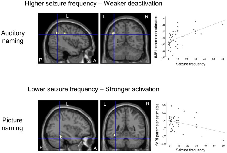
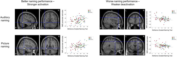
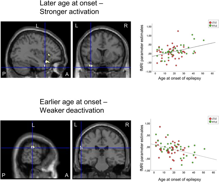
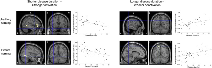
Auditory and picture naming fMRI activated the left posterior temporal lobe, and stronger activation correlated with better clinical naming scores. Verbal fluency MRI mainly activated frontal lobe regions. In left and right TLE, a later age of epilepsy onset related to stronger temporal lobe activations, while earlier age of onset was associated with impaired deactivation of extratemporal regions. In left TLE patients, longer disease duration and higher seizure frequency were associated with reduced deactivation. Frontal lobe language networks were unaffected by disease characteristics.
While frontal lobe language regions appear spared, temporal lobe language areas are susceptible to dysfunction and reorganisation, particularly in left TLE. Early onset and long duration of epilepsy, and high seizure frequency, were associated with compromised activation and deactivation patterns of task-associated regions, which might account for impaired naming performance in individuals with TLE.
利用功能磁共振成像(fMRI)研究颞叶癫痫(TLE)患者语言网络的改变及其与命名障碍的关系。
对 72 例成年 TLE 患者(41 例左侧)和 36 名对照者进行了显性听觉和图片命名 fMRI 任务,以评估颞叶语言区,并进行了隐性言语流畅性任务,以探查额叶语言区。采用全脑多元回归分析,研究 fMRI 激活与临床命名评分的相关性,以及语言网络模式的改变与癫痫持续时间、发病年龄和发作频率的关系。
听觉和图片命名 fMRI 激活了左侧后颞叶,更强的激活与更好的临床命名评分相关。言语流畅性 MRI 主要激活了额叶区域。在左、右 TLE 中,癫痫发病年龄较晚与颞叶激活增强有关,而发病年龄较早与颞外区域去激活受损有关。在左 TLE 患者中,疾病持续时间较长和发作频率较高与去激活减少有关。疾病特征与额叶语言网络无相关性。
虽然额叶语言区域似乎未受影响,但颞叶语言区易发生功能障碍和重组,特别是在左 TLE 中。癫痫的早期发病和较长的病程以及较高的发作频率与任务相关区域的激活和去激活模式受损有关,这可能是 TLE 患者命名障碍的原因。