Mirza-Aghazadeh-Attari Mohammad, Zarrintan Armin, Nezami Nariman, Mohammadi Afshin, Zarrintan Anita, Mohebbi Iraj, Pirnejad Habibollah, Khademvatani Kamal, Ashkavand Zahra, Forughi Payman, Arasteh Amin, Attari Javad Aghazadeh
Aging Research Institute, Tabriz University of Medical Sciences, Tabriz, Iran.
Medical Radiation Research Center, Tabriz University of Medical Sciences, Tabriz, Iran.
Emerg Radiol. 2020 Dec;27(6):653-661. doi: 10.1007/s10140-020-01833-x. Epub 2020 Aug 8.
Computed tomography (CT) has been utilized as a diagnostic modality in the coronavirus disease 19 (COVID-19), while some studies have also suggested a prognostic role for it. This study aimed to assess the diagnostic and prognostic value of computed tomography (CT) imaging in COVID-19 patients.
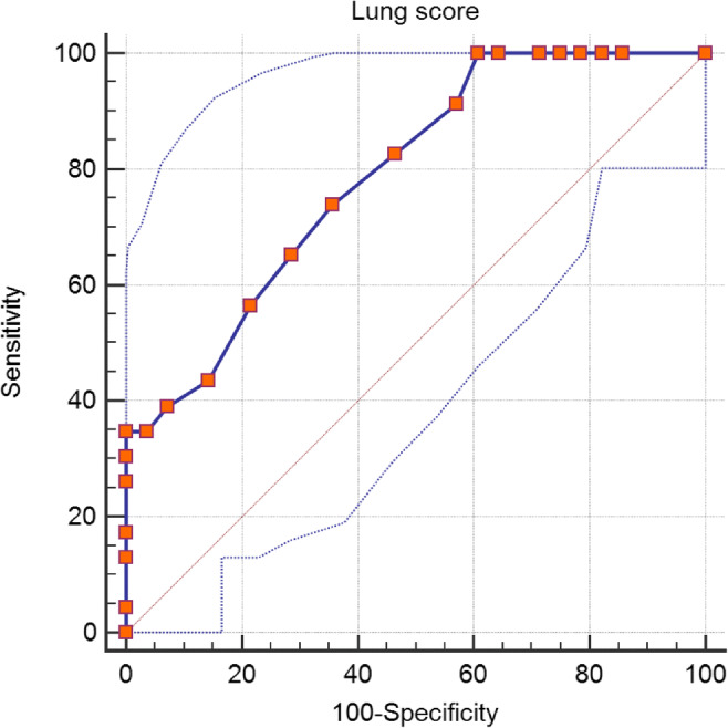
This was a retrospective study of fifty patients with COVID-19 pneumonia. Twenty-seven patients survived, while 23 passed away. CT imaging was performed in all of the patients on the day of admission. Imaging findings were interpreted based on current guidelines by two expert radiologists. Imaging findings were compared between surviving and deceased patients. Lung scores were assigned to patients based on CT chest findings. Then, the receiver operating characteristic curve was used to determine cutoff values for lung scores.
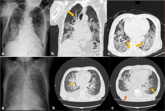
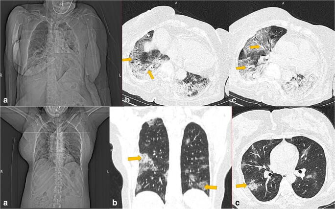
The common radiologic findings were ground-glass opacities (82%) and airspace consolidation (42%), respectively. Air bronchogram was more commonly seen in deceased patients (p = 0.04). Bilateral and multilobar involvement was more frequently found in deceased patients (p = 0.049 and 0.014, respectively). The mean number of involved lobes was 3.46 ± 1.80 lobes in surviving patients and 4.57 ± 0.60 lobes in the deceased patients (p = 0.009). The difference was statistically significant. The area under the curve for a lung score cutoff of 12 was 0.790.
Air bronchogram and bilateral and multilobar involvement were more frequently seen in deceased patients and may suggest a poor outcome for COVID-19 pneumonia.
计算机断层扫描(CT)已被用作冠状病毒病19(COVID-19)的诊断手段,而一些研究也表明其具有预后作用。本研究旨在评估CT成像在COVID-19患者中的诊断和预后价值。
这是一项对50例COVID-19肺炎患者的回顾性研究。27例患者存活,23例死亡。所有患者在入院当天均进行了CT成像。两名专家放射科医生根据现行指南对影像结果进行解读。比较存活患者和死亡患者的影像结果。根据胸部CT表现为患者分配肺部评分。然后,使用受试者操作特征曲线确定肺部评分的临界值。
常见的放射学表现分别为磨玻璃影(82%)和实变影(42%)。空气支气管征在死亡患者中更常见(p = 0.04)。双侧和多叶受累在死亡患者中更常见(分别为p = 0.049和0.014)。存活患者受累肺叶的平均数量为3.46±1.80个肺叶,死亡患者为4.57±0.60个肺叶(p = 0.009)。差异具有统计学意义。肺部评分临界值为12时的曲线下面积为0.790。
空气支气管征以及双侧和多叶受累在死亡患者中更常见,可能提示COVID-19肺炎预后不良。