Paletta George A, Crane David M, Konicek John, Piepenbrink Marina, Higgins Laurence D, Milner John D, Wijdicks Coen A
The Orthopedic Center of St Louis, Chesterfield, Missouri, USA.
Bluetail Medical Group, St Louis, Missouri, USA.
Orthop J Sports Med. 2020 Jul 29;8(7):2325967120936672. doi: 10.1177/2325967120936672. eCollection 2020 Jul.
Meniscal extrusion refers to meniscal displacement out of the joint space and over the tibial margin, altering knee mechanics and increasing the risk of osteoarthritis. The meniscotibial ligaments have been shown to have an important role in meniscal stability. However, it remains unclear whether an isolated lesion of the medial meniscotibial ligaments will result in meniscal extrusion and whether repairing the detached ligament will reduce extrusion.
A lesion of the medial meniscotibial ligament will result in meniscal extrusion, and repairing the joint capsule will eliminate the extrusion by returning the meniscus back to its original position.
Controlled laboratory study.
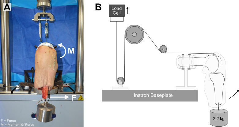
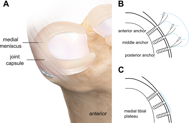
Fresh-frozen human cadaveric knees (N = 6) were used for biomechanical testing. The test protocol involved 100 flexion-extension cycles. In full extension, meniscal extrusion was measured using ultrasound, in both an otherwise unloaded state and while subjected to a 10-N·m varus load. Each knee was tested in its native condition (baseline), after creating a detachment of the medial meniscotibial ligament, and finally with the joint capsule repaired using 3 knotless SutureTak anchors. We also performed a retrospective review of 15 patients who underwent meniscotibial ligament repair with a minimal follow-up of 5 weeks (mean, 14 weeks; range, 5-35 weeks).
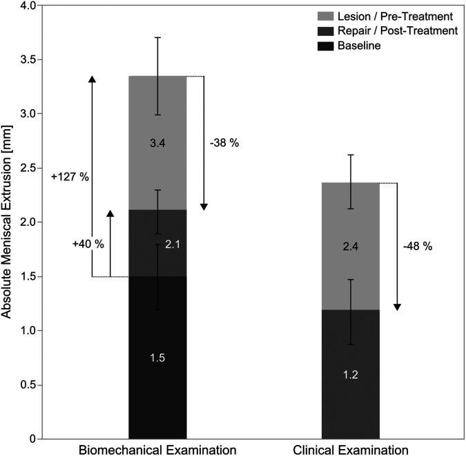
During biomechanical testing, the mean absolute meniscal extrusion at baseline was 1.5 ± 0.6 mm. After creation of the meniscotibial ligament lesion, the mean absolute meniscal extrusion was significantly increased (3.4 ± 0.7 mm) ( < .001). After repair, the extrusion was reduced to 2.1 ± 0.4 mm ( < .001). Clinically, a reduction in absolute meniscal extrusion of approximately 48% was reached (1.2 ± 0.6 vs 2.4 ± 0.5 mm preoperatively; < .001).
This study indicates that the medial meniscotibial ligaments contribute to meniscal stability as lesions cause the meniscus to extrude and that repair of those ligaments can significantly reduce extrusion. Early clinical results using this meniscotibial ligament repair technique support our biomechanical findings, as a significant reduction in meniscal extrusion was achieved.
Our biomechanical findings suggest that repair of medial meniscotibial ligaments reduces meniscal extrusion and clinically may improve meniscal function, with the possible long-term benefit of reducing the risk for osteoarthritis.
半月板挤出是指半月板移位出关节间隙并越过胫骨边缘,改变膝关节力学并增加骨关节炎风险。半月板胫骨韧带已被证明在半月板稳定性中起重要作用。然而,内侧半月板胫骨韧带的孤立损伤是否会导致半月板挤出以及修复分离的韧带是否会减少挤出仍不清楚。
内侧半月板胫骨韧带损伤会导致半月板挤出,修复关节囊将通过使半月板恢复到其原始位置来消除挤出。
对照实验室研究。
使用新鲜冷冻的人体尸体膝关节(N = 6)进行生物力学测试。测试方案包括100次屈伸循环。在完全伸展时,在未加载状态和承受10 N·m内翻负荷时,使用超声测量半月板挤出。每个膝关节在其原始状态(基线)、内侧半月板胫骨韧带分离后以及最后使用3个无结SutureTak锚钉修复关节囊后进行测试。我们还对15例接受半月板胫骨韧带修复的患者进行了回顾性研究,最短随访5周(平均14周;范围5 - 35周)。
在生物力学测试期间,基线时半月板平均绝对挤出为1.5±0.6 mm。内侧半月板胫骨韧带损伤后,半月板平均绝对挤出显著增加(3.4±0.7 mm)(P <.001)。修复后,挤出减少至2.1±0.4 mm(P <.001)。临床上,半月板绝对挤出减少约48%(术前为1.2±0.6 vs 2.4±0.5 mm;P <.001)。
本研究表明,内侧半月板胫骨韧带有助于半月板稳定性,因为损伤会导致半月板挤出,并且修复这些韧带可显著减少挤出。使用这种半月板胫骨韧带修复技术的早期临床结果支持我们的生物力学研究结果,因为半月板挤出显著减少。
我们的生物力学研究结果表明,内侧半月板胫骨韧带的修复可减少半月板挤出,临床上可能改善半月板功能,可能具有降低骨关节炎风险的长期益处。