Camstra Kevin M, Srinivasan Visish M, Collins Dalis, Chen Stephen, Kan Peter, Johnson Jeremiah
Department of Neurosurgery, Baylor College of Medicine, Houston, TX, USA.
Department of Surgery, Baylor College of Medicine, Houston, TX, USA.
Neurointervention. 2020 Nov;15(3):107-116. doi: 10.5469/neuroint.2020.00150. Epub 2020 Aug 11.
With advancing endovascular technology and increasing interest in minimally invasive intra-arterial therapies such as stem cell and chemotherapy for cerebral disease, the establishment of a translational model with cerebral circulation accessible to microcatheters is needed. We report our experience catheterizing canine cerebral circulation with microcatheters, present high-resolution angiographic images of the canine vascular anatomy, describe arterial branch flow patterns and provide measurements of canine arterial conduits.
Angiograms were performed on 10 intact purpose-bred hounds. Angiography, measurements of arterial conduits and catheterization information for intracranial arterial branches were obtained.
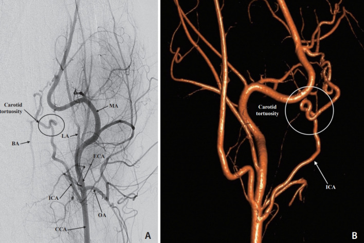
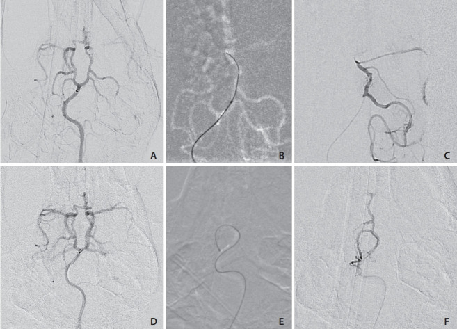
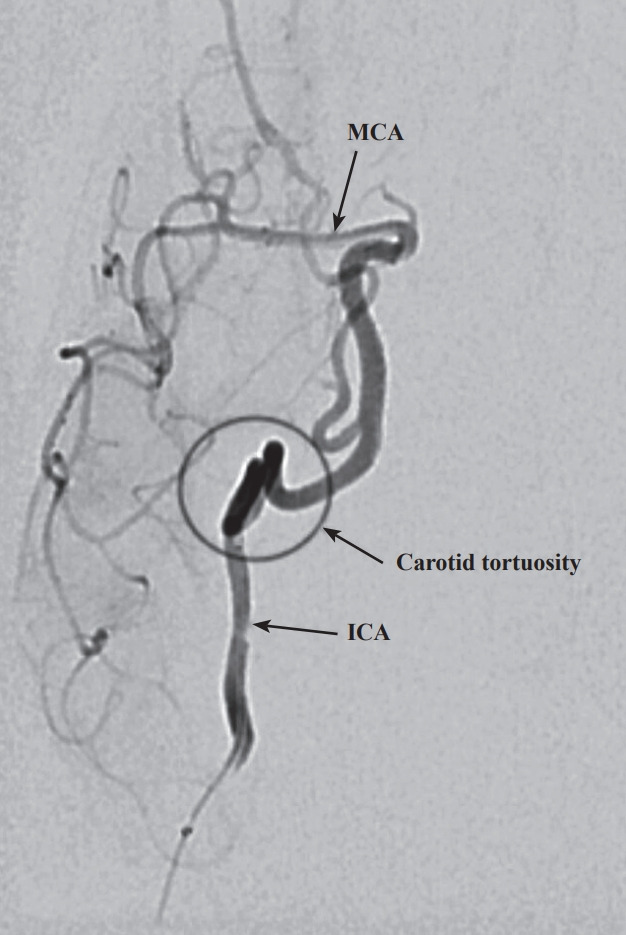
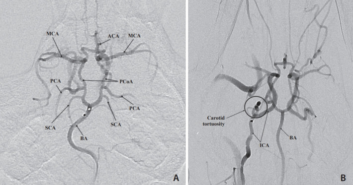
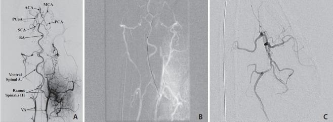
Selective and superselective cerebral angiography was successful in all subjects. Relevant arterial mean diameters include the femoral (4.64 mm), aorta (9.38 mm), external carotid (3.65 mm), internal carotid arteries (1.6 mm), vertebrobasilar system and Circle of Willis branches. Catheterization of the Circle of Willis was achieved via the posterior circulation in all subjects tested (n=3) and the use of flow directed microcatheters resulted in reduced arterial tree deformation and improved superselection of intracranial vessels. Catheterization of the intracranial circulation was attempted but not achieved via the internal carotid artery (n=7) due to its tortuosity and subsequent catheter related vasospasm.
The canine cerebral vasculature is posterior circulation dominant. Anterior circulation angiography is achievable via the internal carotid artery, but direct cerebral arterial access is best achieved via the posterior circulation using flow-directed microcatheters. It is feasible to deliver intra-arterial therapies to selective vascular territories within the canine cerebral circulation, thus making it a viable animal model for testing novel intra-arterial cerebral treatments.
随着血管内技术的进步以及对微创动脉内治疗(如用于脑部疾病的干细胞和化疗)的兴趣日益增加,需要建立一种可通过微导管进入脑循环的转化模型。我们报告了使用微导管对犬脑循环进行插管的经验,展示了犬血管解剖结构的高分辨率血管造影图像,描述了动脉分支血流模式并提供了犬动脉管道的测量数据。
对10只完整的纯种猎犬进行血管造影。获得了血管造影、动脉管道测量以及颅内动脉分支的插管信息。
所有受试者均成功进行了选择性和超选择性脑血管造影。相关动脉平均直径包括股动脉(4.64毫米)、主动脉(9.38毫米)、颈外动脉(3.65毫米)、颈内动脉(1.6毫米)、椎基底系统和 Willis 环分支。在所有测试的受试者(n = 3)中,通过后循环实现了 Willis 环的插管,使用血流导向微导管可减少动脉树变形并改善颅内血管的超选择性。由于颈内动脉迂曲以及随后与导管相关的血管痉挛,尝试通过颈内动脉对颅内循环进行插管但未成功(n = 7)。
犬脑循环系统以后循环为主。通过颈内动脉可实现前循环血管造影,但使用血流导向微导管通过后循环进行直接脑动脉穿刺效果最佳。将动脉内治疗输送到犬脑循环内的选择性血管区域是可行的,因此使其成为测试新型动脉内脑治疗方法的可行动物模型。