Zhang Yunfeng, Jin Min, Du Bin, Lin Hao, Xu Chengyong, Jiang Weijian, Jia Jianping
Department of Neurology, Xuan Wu Hospital, Capital Medical University, Beijing, China.
Department of Neurology, Affiliated Hospital of Nantong University, Nantong, China.
PLoS One. 2015 Nov 6;10(11):e0142251. doi: 10.1371/journal.pone.0142251. eCollection 2015.
The extended time window and theoretic reduction in hemorrhage make mechanical strategies an attractive approach for the treatment of patients with ischemic stroke. However, a limited availability of suitable animal models of cerebrovascular thrombosis has hampered the study of novel endovascular interventions. The aim of the present study was to develop a new technique for site-specific placement of a thrombus in a canine model that would allow for the evaluation of mechanical thrombectomy and clot retrieval methods and the visualization of thrombus dislocation or fragmentation during angiographic manipulation.
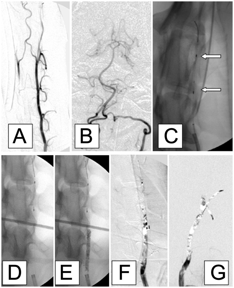
Angiography and embolization with a preformed thrombus were performed in 12 canines. Under fluoroscopic guidance, an embolism protection device (EPD) was anchored to the middle segment of the left vertebral artery (VA) via the left femoral arterial sheath. A preformed radiopaque clot was injected through the guide catheter into the left VA, via the contralateral femoral artery, proximal to the EPD. After 15 min of occlusion, the EPD was removed and persistent occlusion of the VA was documented angiographically.
Angiography performed during the observation period confirmed the persistence of VA occlusion in each case, and displacement of the radiopaque clots did not occur during the 3-hour observation period. The technique allowed selective embolization of targeted vessels without thrombus fragmentation.
This study demonstrates, for the first time, a canine model of post-circulation embolism induced by autologous blood clot placement. This model can be rapidly formed and easily operated, and the site of thrombosis can be readily controlled.
延长的时间窗以及理论上出血风险的降低使机械治疗策略成为缺血性脑卒中患者治疗的一种有吸引力的方法。然而,合适的脑血管血栓形成动物模型数量有限,阻碍了新型血管内介入治疗的研究。本研究的目的是开发一种新技术,在犬模型中进行血栓的特定部位放置,以评估机械取栓和血栓清除方法,并在血管造影操作过程中观察血栓移位或破碎情况。
对12只犬进行血管造影和预制血栓栓塞术。在荧光透视引导下,通过左股动脉鞘将栓塞保护装置(EPD)固定于左椎动脉(VA)中段。通过引导导管经对侧股动脉在EPD近端将预制的不透射线血栓注入左VA。闭塞15分钟后,移除EPD,并通过血管造影记录VA的持续闭塞情况。
观察期内进行的血管造影证实每例VA均持续闭塞,在3小时观察期内不透射线血栓未发生移位。该技术可实现目标血管的选择性栓塞且无血栓破碎。
本研究首次证明了一种通过自体血凝块放置诱导后循环栓塞的犬模型。该模型形成迅速、操作简便,血栓形成部位易于控制。