Department of Otolaryngology, University of Pittsburgh Medical Center, Pittsburgh, Pennsylvania, USA.
University of Pittsburgh School of Medicine, University of Pittsburgh, Pittsburgh, Pennsylvania, USA.
Otolaryngol Head Neck Surg. 2021 Feb;164(2):285-293. doi: 10.1177/0194599820949802. Epub 2020 Aug 11.
To define the aerosol and droplet risks associated with endonasal drilling and to identify mitigation strategies.
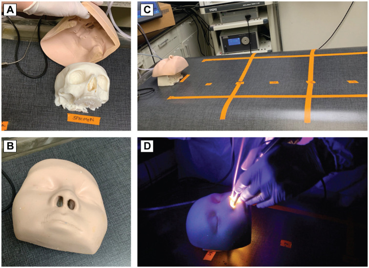
Simulation series with fluorescent 3-dimensional (3D) printed sinonasal models and deidentified cadaveric heads.
Dedicated surgical laboratory.
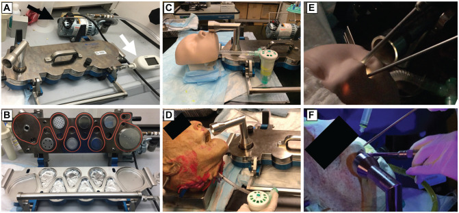
Cadaveric specimens irrigated with fluorescent tracer and fluorescent 3D-printed models were drilled. A cascade impactor was used to collect aerosols and small droplets of various aerodynamic diameters under 15 µm. Large droplet generation was measured by evaluating the field for fluorescent debris. Aerosol plumes through the nares were generated via nebulizer, and mitigation measures, including suction and SPIWay devices, nasal sheaths, were evaluated regarding reduction of aerosol escape from the nose.
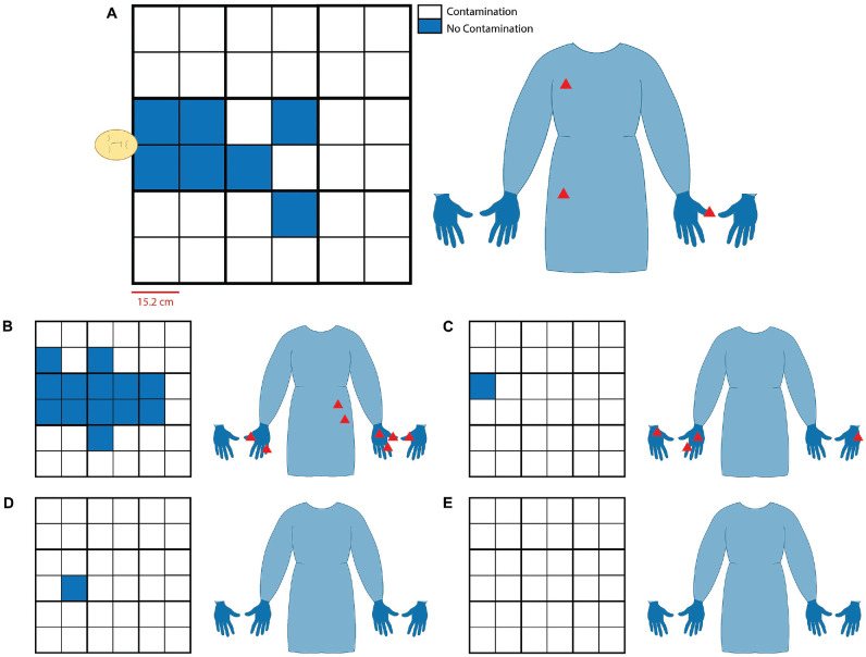
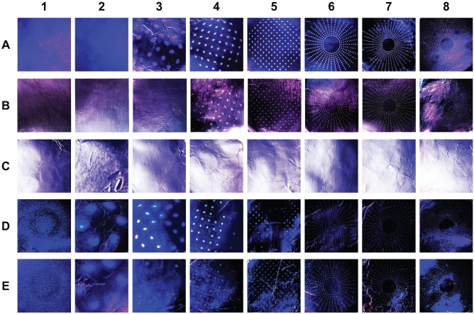
The drilling of cadaveric specimens without flexible suction generated aerosols ≤3.30 µm, and drilling of 3D sinonasal models consistently produced aerosols ≤14.1 µm. Mitigation with SPIWay or diameter-restricted SPIWay produced same results. There was minimal field contamination in the cadaveric models, 0% to 2.77% field tarp area, regardless of drill burr type or drilling location; cutting burr drilling without suction in the 3D model yielded the worst contamination field (36.1%), followed by coarse diamond drilling without suction (19.4%). The simple placement of a flexible suction instrument in the nasal cavity or nasopharynx led to complete elimination of all aerosols ≤14.1 µm, as evaluated by a cascade impactor positioned immediately at the nares.
Given the findings regarding aerosol risk reduction, we strongly recommend that physicians use a suction instrument in the nasal cavity or nasopharynx during endonasal surgery in the COVID-19 era.
定义经鼻内钻孔相关的气溶胶和液滴风险,并确定减轻风险的策略。
使用荧光 3D 打印鼻窦模型和去识别尸体头部的模拟系列。
专用手术实验室。
用荧光示踪剂和荧光 3D 打印模型冲洗尸体标本,并进行钻孔。使用级联撞击器收集各种空气动力学直径小于 15 µm 的气溶胶和小液滴。通过评估荧光碎片的区域来测量大液滴的产生。通过喷雾器产生经鼻腔的气溶胶羽流,并评估各种吸气和 SPIWay 装置、鼻腔护套等减轻措施,以减少从鼻腔逸出的气溶胶。
在没有柔性抽吸的情况下对尸体标本进行钻孔会产生 ≤3.30 µm 的气溶胶,而对 3D 鼻窦模型进行钻孔则会持续产生 ≤14.1 µm 的气溶胶。使用 SPIWay 或限制直径的 SPIWay 进行减轻措施会产生相同的结果。尸体模型中的污染区域最小,为 0%至 2.77%的场篷面积,无论钻头类型或钻孔位置如何;在 3D 模型中无抽吸的切割钻头会产生最严重的污染场(36.1%),其次是无抽吸的粗金刚石钻头(19.4%)。简单地将柔性抽吸仪器放置在鼻腔或鼻咽中,通过立即放置在鼻腔处的级联撞击器,可完全消除所有 ≤14.1 µm 的气溶胶。
鉴于气溶胶风险降低的发现,我们强烈建议在 COVID-19 时代,医生在经鼻内手术期间使用鼻腔或鼻咽中的抽吸仪器。