• 文献检索
  • 文档翻译
  • 深度研究
  • 学术资讯
  • Suppr Zotero 插件Zotero 插件
  • 邀请有礼
  • 套餐&价格
  • 历史记录
应用&插件
Suppr Zotero 插件Zotero 插件浏览器插件Mac 客户端Windows 客户端微信小程序
定价
高级版会员购买积分包购买API积分包
服务
文献检索文档翻译深度研究API 文档MCP 服务
关于我们
关于 Suppr公司介绍联系我们用户协议隐私条款
关注我们

Suppr 超能文献

核心技术专利:CN118964589B侵权必究
粤ICP备2023148730 号-1Suppr @ 2026

文献检索

告别复杂PubMed语法,用中文像聊天一样搜索,搜遍4000万医学文献。AI智能推荐,让科研检索更轻松。

立即免费搜索

文件翻译

保留排版,准确专业,支持PDF/Word/PPT等文件格式,支持 12+语言互译。

免费翻译文档

深度研究

AI帮你快速写综述,25分钟生成高质量综述,智能提取关键信息,辅助科研写作。

立即免费体验

结合多视图二维卷积神经网络的主动脉计算机断层扫描血管造影的3D自动分割

3D Automatic Segmentation of Aortic Computed Tomography Angiography Combining Multi-View 2D Convolutional Neural Networks.

作者信息

Fantazzini Alice, Esposito Mario, Finotello Alice, Auricchio Ferdinando, Pane Bianca, Basso Curzio, Spinella Giovanni, Conti Michele

机构信息

Department of Experimental Medicine, University of Genoa, Via Leon Battista Alberti, 2, 16132, Genoa, Italy.

Camelot Biomedical Systems S.r.l, Via Al Ponte Reale, 2, 16124, Genoa, Italy.

出版信息

Cardiovasc Eng Technol. 2020 Oct;11(5):576-586. doi: 10.1007/s13239-020-00481-z. Epub 2020 Aug 11.

DOI:10.1007/s13239-020-00481-z
PMID:32783134
原文链接:https://pmc.ncbi.nlm.nih.gov/articles/PMC7511465/
Abstract

PURPOSE

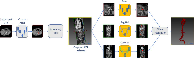
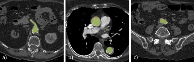
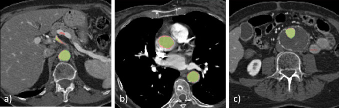
The quantitative analysis of contrast-enhanced Computed Tomography Angiography (CTA) is essential to assess aortic anatomy, identify pathologies, and perform preoperative planning in vascular surgery. To overcome the limitations given by manual and semi-automatic segmentation tools, we apply a deep learning-based pipeline to automatically segment the CTA scans of the aortic lumen, from the ascending aorta to the iliac arteries, accounting for 3D spatial coherence.

METHODS

A first convolutional neural network (CNN) is used to coarsely segment and locate the aorta in the whole sub-sampled CTA volume, then three single-view CNNs are used to effectively segment the aortic lumen from axial, sagittal, and coronal planes under higher resolution. Finally, the predictions of the three orthogonal networks are integrated to obtain a segmentation with spatial coherence.

RESULTS

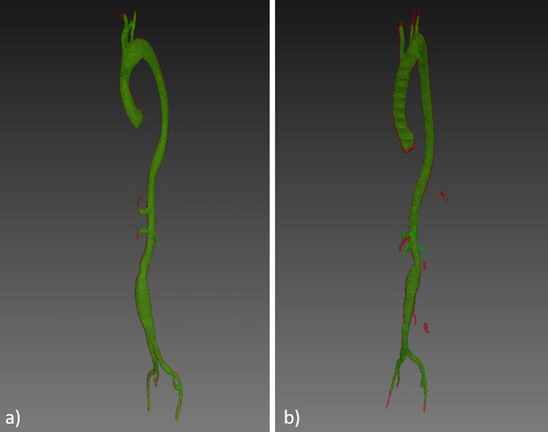
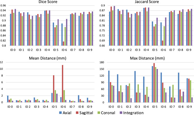
The coarse segmentation performed to identify the aortic lumen achieved a Dice coefficient (DSC) of 0.92 ± 0.01. Single-view axial, sagittal, and coronal CNNs provided a DSC of 0.92 ± 0.02, 0.92 ± 0.04, and 0.91 ± 0.02, respectively. Multi-view integration provided a DSC of 0.93 ± 0.02 and an average surface distance of 0.80 ± 0.26 mm on a test set of 10 CTA scans. The generation of the ground truth dataset took about 150 h and the overall training process took 18 h. In prediction phase, the adopted pipeline takes around 25 ± 1 s to get the final segmentation.

CONCLUSION

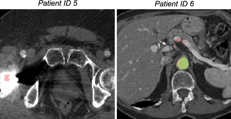
The achieved results show that the proposed pipeline can effectively localize and segment the aortic lumen in subjects with aneurysm.

摘要

目的

对比增强计算机断层血管造影(CTA)的定量分析对于评估主动脉解剖结构、识别病变以及在血管外科手术中进行术前规划至关重要。为了克服手动和半自动分割工具的局限性,我们应用基于深度学习的流程来自动分割从升主动脉到髂动脉的主动脉腔CTA扫描图像,同时考虑三维空间连贯性。

方法

首先使用一个卷积神经网络(CNN)在整个下采样的CTA体积中对主动脉进行粗略分割和定位,然后使用三个单视图CNN在更高分辨率下从轴向、矢状和冠状平面有效地分割主动脉腔。最后,将三个正交网络的预测结果进行整合,以获得具有空间连贯性的分割结果。

结果

用于识别主动脉腔的粗略分割的骰子系数(DSC)为0.92±0.01。单视图轴向、矢状和冠状CNN的DSC分别为0.92±0.02、0.92±0.04和0.91±0.02。在一个包含10次CTA扫描的测试集上,多视图整合的DSC为0.93±0.02,平均表面距离为0.80±0.26毫米。生成地面真值数据集耗时约150小时,整个训练过程耗时18小时。在预测阶段,所采用的流程大约需要25±1秒来获得最终分割结果。

结论

所取得的结果表明,所提出的流程能够有效地定位和分割患有动脉瘤患者的主动脉腔。

https://cdn.ncbi.nlm.nih.gov/pmc/blobs/3b7f/7511465/3ccac944a3fe/13239_2020_481_Fig6_HTML.jpg
https://cdn.ncbi.nlm.nih.gov/pmc/blobs/3b7f/7511465/1c9a04b94b85/13239_2020_481_Fig1_HTML.jpg
https://cdn.ncbi.nlm.nih.gov/pmc/blobs/3b7f/7511465/53201f1a99a5/13239_2020_481_Fig2_HTML.jpg
https://cdn.ncbi.nlm.nih.gov/pmc/blobs/3b7f/7511465/2fc1a1dc0e30/13239_2020_481_Fig3_HTML.jpg
https://cdn.ncbi.nlm.nih.gov/pmc/blobs/3b7f/7511465/f84b899edfda/13239_2020_481_Fig4_HTML.jpg
https://cdn.ncbi.nlm.nih.gov/pmc/blobs/3b7f/7511465/7120f6b4ef22/13239_2020_481_Fig5_HTML.jpg
https://cdn.ncbi.nlm.nih.gov/pmc/blobs/3b7f/7511465/3ccac944a3fe/13239_2020_481_Fig6_HTML.jpg

相似文献

1
3D Automatic Segmentation of Aortic Computed Tomography Angiography Combining Multi-View 2D Convolutional Neural Networks.结合多视图二维卷积神经网络的主动脉计算机断层扫描血管造影的3D自动分割
Cardiovasc Eng Technol. 2020 Oct;11(5):576-586. doi: 10.1007/s13239-020-00481-z. Epub 2020 Aug 11.
2
Deep Learning to Automatically Segment and Analyze Abdominal Aortic Aneurysm from Computed Tomography Angiography.深度学习自动分割和分析计算机断层血管造影中的腹主动脉瘤。
Cardiovasc Eng Technol. 2022 Aug;13(4):535-547. doi: 10.1007/s13239-021-00594-z. Epub 2022 Jan 8.
3
Deep learning-aided extraction of outer aortic surface from CT angiography scans of patients with Stanford type B aortic dissection.深度学习辅助从 Stanford 型 B 主动脉夹层患者的 CT 血管造影扫描中提取主动脉外表面。
Eur Radiol Exp. 2023 Jun 29;7(1):35. doi: 10.1186/s41747-023-00342-z.
4
Automatic Segmentation, Detection, and Diagnosis of Abdominal Aortic Aneurysm (AAA) Using Convolutional Neural Networks and Hough Circles Algorithm.使用卷积神经网络和霍夫圆算法对腹主动脉瘤(AAA)进行自动分割、检测和诊断
Cardiovasc Eng Technol. 2019 Sep;10(3):490-499. doi: 10.1007/s13239-019-00421-6. Epub 2019 Jun 19.
5
Multicentric clinical evaluation of a computed tomography-based fully automated deep neural network for aortic maximum diameter and volumetric measurements.基于计算机断层扫描的主动脉最大直径和容积测量全自动深度神经网络的多中心临床评估。
J Vasc Surg. 2024 Jun;79(6):1390-1400.e8. doi: 10.1016/j.jvs.2024.01.214. Epub 2024 Feb 5.
6
Fully automatic volume segmentation of infrarenal abdominal aortic aneurysm computed tomography images with deep learning approaches versus physician controlled manual segmentation.基于深度学习的自动与医生手动勾画肾下腹部主动脉瘤 CT 图像容积比较。
J Vasc Surg. 2021 Jul;74(1):246-256.e6. doi: 10.1016/j.jvs.2020.11.036. Epub 2020 Dec 9.
7
Fully automatic segmentation of type B aortic dissection from CTA images enabled by deep learning.基于深度学习的 CTA 图像中 B 型主动脉夹层的全自动分割。
Eur J Radiol. 2019 Dec;121:108713. doi: 10.1016/j.ejrad.2019.108713. Epub 2019 Oct 17.
8
Pre-surgical and Post-surgical Aortic Aneurysm Maximum Diameter Measurement: Full Automation by Artificial Intelligence.术前和术后主动脉瘤最大直径测量:人工智能全自动测量。
Eur J Vasc Endovasc Surg. 2021 Dec;62(6):869-877. doi: 10.1016/j.ejvs.2021.07.013. Epub 2021 Sep 10.
9
Fully automatic segmentation of abdominal aortic thrombus in pre-operative CTA images using deep convolutional neural networks.使用深度卷积神经网络对术前 CTA 图像中的腹部主动脉血栓进行全自动分割。
Technol Health Care. 2022;30(5):1257-1266. doi: 10.3233/THC-THC213630.
10
Fully automatic detection and segmentation of abdominal aortic thrombus in post-operative CTA images using Deep Convolutional Neural Networks.使用深度卷积神经网络对术后 CTA 图像中的腹部主动脉血栓进行全自动检测和分割。
Med Image Anal. 2018 May;46:202-214. doi: 10.1016/j.media.2018.03.010. Epub 2018 Mar 27.

引用本文的文献

1
Automatic CTA analysis for blood vessels and aneurysm features extraction in EVAR planning.用于血管内腹主动脉瘤修复(EVAR)规划中血管和动脉瘤特征提取的自动CT血管造影(CTA)分析
Sci Rep. 2025 May 12;15(1):16431. doi: 10.1038/s41598-025-00484-7.
2
Application of three-dimensional printing in the planning and execution of aortic aneurysm repair.三维打印在主动脉瘤修复手术规划与实施中的应用。
Front Cardiovasc Med. 2025 Jan 29;11:1485267. doi: 10.3389/fcvm.2024.1485267. eCollection 2024.
3
Assessment of the Aorto-Septal Angle Post-Thoracic Endovascular Aortic Repair through Segmentation and the Semi-Automatic Analysis of Cardiosynchronized Computed Tomography Angiography Images.

本文引用的文献

1
Abdominal Aortic Aneurysm Segmentation Using Convolutional Neural Networks Trained with Images Generated with a Synthetic Shape Model.使用基于合成形状模型生成的图像训练的卷积神经网络进行腹主动脉瘤分割
Mach Learn Med Eng Cardiovasc Health Intravasc Imaging Comput Assist Stenting (2019). 2019;11794:167-174. doi: 10.1007/978-3-030-33327-0_20. Epub 2019 Oct 12.
2
Fully automatic segmentation of type B aortic dissection from CTA images enabled by deep learning.基于深度学习的 CTA 图像中 B 型主动脉夹层的全自动分割。
Eur J Radiol. 2019 Dec;121:108713. doi: 10.1016/j.ejrad.2019.108713. Epub 2019 Oct 17.
3
通过对心脏同步计算机断层扫描血管造影图像进行分割和半自动分析来评估胸主动脉腔内修复术后的主动脉-室间隔角
J Cardiovasc Dev Dis. 2024 Sep 4;11(9):275. doi: 10.3390/jcdd11090275.
4
Aorta Segmentation in 3D CT Images by Combining Image Processing and Machine Learning Techniques.结合图像处理与机器学习技术的三维CT图像主动脉分割
Cardiovasc Eng Technol. 2024 Jun;15(3):359-373. doi: 10.1007/s13239-024-00720-7. Epub 2024 Feb 22.
5
Machine learning-based identification of contrast-enhancement phase of computed tomography scans.基于机器学习的计算机断层扫描增强扫描相的识别。
PLoS One. 2024 Feb 2;19(2):e0294581. doi: 10.1371/journal.pone.0294581. eCollection 2024.
6
Artificial Intelligence Application to Screen Abdominal Aortic Aneurysm Using Computed tomography Angiography.人工智能应用于计算机断层血管造影筛查腹主动脉瘤。
J Digit Imaging. 2023 Oct;36(5):2125-2137. doi: 10.1007/s10278-023-00866-1. Epub 2023 Jul 5.
7
Deep-learning method for fully automatic segmentation of the abdominal aortic aneurysm from computed tomography imaging.基于计算机断层扫描成像的腹主动脉瘤全自动分割深度学习方法。
Front Cardiovasc Med. 2023 Jan 5;9:1040053. doi: 10.3389/fcvm.2022.1040053. eCollection 2022.
8
3D modeling and printing for complex biventricular repair of double outlet right ventricle.用于右心室双出口复杂双心室修复的3D建模与打印
Front Cardiovasc Med. 2022 Nov 30;9:1024053. doi: 10.3389/fcvm.2022.1024053. eCollection 2022.
9
A fully automated deep learning pipeline for micro-CT-imaging-based densitometry of lung fibrosis murine models.一种基于深度学习的全自动微 CT 成像肺纤维化小鼠模型骨密度分析的方法。
Respir Res. 2022 Nov 11;23(1):308. doi: 10.1186/s12931-022-02236-x.
10
Fast Approximate Quantification of Endovascular Stent Graft Displacement Forces in the Bovine Aortic Arch Variant.快速近似量化牛主动脉弓变异模型中血管内支架移植物的置换力。
J Endovasc Ther. 2023 Oct;30(5):756-768. doi: 10.1177/15266028221095403. Epub 2022 May 19.
A fully automated pipeline for mining abdominal aortic aneurysm using image segmentation.
基于图像分割的全自动挖掘腹主动脉瘤的流水线。
Sci Rep. 2019 Sep 24;9(1):13750. doi: 10.1038/s41598-019-50251-8.
4
Automatic Segmentation, Detection, and Diagnosis of Abdominal Aortic Aneurysm (AAA) Using Convolutional Neural Networks and Hough Circles Algorithm.使用卷积神经网络和霍夫圆算法对腹主动脉瘤(AAA)进行自动分割、检测和诊断
Cardiovasc Eng Technol. 2019 Sep;10(3):490-499. doi: 10.1007/s13239-019-00421-6. Epub 2019 Jun 19.
5
QuickNAT: A fully convolutional network for quick and accurate segmentation of neuroanatomy.QuickNAT:用于快速准确分割神经解剖结构的全卷积网络。
Neuroimage. 2019 Feb 1;186:713-727. doi: 10.1016/j.neuroimage.2018.11.042. Epub 2018 Nov 29.
6
Automated nasopharyngeal carcinoma segmentation in magnetic resonance images by combination of convolutional neural networks and graph cut.通过卷积神经网络与图割相结合实现磁共振图像中鼻咽癌的自动分割
Exp Ther Med. 2018 Sep;16(3):2511-2521. doi: 10.3892/etm.2018.6478. Epub 2018 Jul 18.
7
Fully automatic detection and segmentation of abdominal aortic thrombus in post-operative CTA images using Deep Convolutional Neural Networks.使用深度卷积神经网络对术后 CTA 图像中的腹部主动脉血栓进行全自动检测和分割。
Med Image Anal. 2018 May;46:202-214. doi: 10.1016/j.media.2018.03.010. Epub 2018 Mar 27.
8
The Society for Vascular Surgery practice guidelines on the care of patients with an abdominal aortic aneurysm.血管外科学会治疗腹主动脉瘤患者的实践指南。
J Vasc Surg. 2018 Jan;67(1):2-77.e2. doi: 10.1016/j.jvs.2017.10.044.
9
Abdominal aortic aneurysm: pictorial review of common appearances and complications.腹主动脉瘤:常见表现及并发症的影像学综述
Ann Transl Med. 2017 Jun;5(12):256. doi: 10.21037/atm.2017.04.32.
10
Deep Learning in Medical Image Analysis.医学图像分析中的深度学习
Annu Rev Biomed Eng. 2017 Jun 21;19:221-248. doi: 10.1146/annurev-bioeng-071516-044442. Epub 2017 Mar 9.